

Glioblastoma, a notoriously aggressive and often fatal form of brain cancer, presents a formidable challenge to medical science. For decades, research has predominantly focused on the genetic mutations and chemical signaling pathways that drive tumor growth and spread. However, a groundbreaking study, published in the prestigious journal Microsystems & Nanoengineering, has illuminated a surprisingly potent factor in glioblastoma’s invasive prowess: the physical properties of the fluid surrounding the tumor. Scientists have discovered that the increasing viscosity of the extracellular fluid at the advancing edges of these tumors, rather than merely impeding their movement, actively compels glioblastoma cells to adapt in ways that enhance their ability to penetrate healthy brain tissue. This research not only challenges long-held assumptions in cancer biology but also offers a promising new avenue for therapeutic development.
The conventional understanding of cancer invasion has largely centered on the biochemical interactions between tumor cells and their microenvironment, alongside the mechanical properties of the surrounding tissue, such as its stiffness. The viscosity of the interstitial fluid—the liquid that bathes cells in tissues—has been largely overlooked as a significant player. This oversight is particularly striking given the dramatic physical gradients that exist within glioblastoma tumors. In a significant departure from fluid found in healthy brain tissue, the fluid at the periphery of glioblastoma is demonstrably more viscous. This study quantifies this difference, revealing that the fluid at the tumor’s advancing edge can be up to eight times thicker than that found in the necrotic, dead tissue at the tumor’s core. This stark contrast creates a progressively challenging physical landscape for migrating cancer cells.
The Challenge of Replicating Tumor Microenvironments
Investigating the precise impact of fluid viscosity on cancer cell behavior has historically been fraught with methodological difficulties. Traditional laboratory models, often relying on static cell cultures or enclosed microfluidic devices, struggle to accurately mimic the dynamic and complex physical conditions experienced by glioblastoma cells in vivo. Enclosed microfluidic systems, while useful for some applications, can introduce confounding factors. The limited access to oxygen and nutrients in such confined spaces can alter cell metabolism and behavior. Furthermore, friction against the device’s artificial walls generates additional forces that can influence cell motility and morphology, making it challenging to isolate the specific effects of fluid viscosity. Crucially, these systems often fall short in their ability to sustain cell cultures for extended periods, thus limiting the observation of long-term adaptive responses to mechanical stress. This inability to accurately replicate and study the prolonged influence of viscous fluid has been a significant impediment to understanding how glioblastoma cells truly adapt and become more invasive.
An Innovative Platform for Unveiling Cellular Adaptations
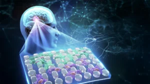
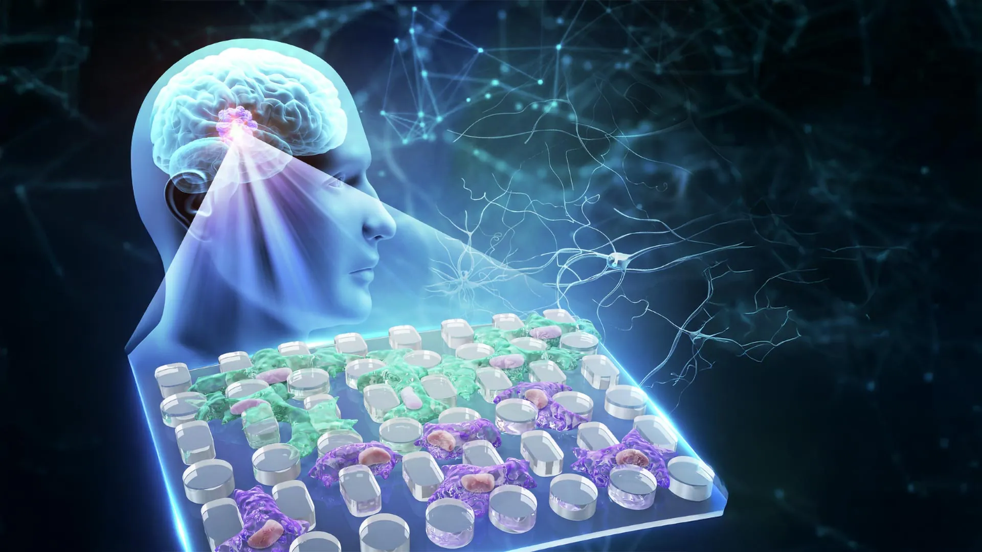
To overcome these limitations, researchers from Chongqing General Hospital and Chongqing University in China have engineered a novel, open microfluidic platform. This innovative device is designed to precisely replicate the physical conditions encountered by glioblastoma cells at the tumor’s invasive front, while simultaneously allowing for extended observation of cellular responses. The platform features a two-layer open microfluidic membrane, equipped with a removable cap and an array of precisely spaced microscopic pillars. This ingenious design allows researchers to meticulously control the initiation of cell migration and observe, in real-time, how cancer cell nuclei change shape as they navigate confined spaces.
A key advantage of this open platform is its capacity for continuous cell culture for up to one month. This extended observation period is critical for understanding how prolonged exposure to mechanical stress, specifically varying fluid viscosity, influences the migratory patterns and invasive capabilities of glioblastoma cells. Unlike closed systems, the open design minimizes extraneous forces and ensures a more consistent supply of essential nutrients and oxygen, creating an environment that more closely mirrors the tumor’s natural setting.
The research team utilized two well-established human glioblastoma cell lines, U-251 and LN-229, for their experiments. These cells were cultured for a full month within the microfluidic platform using a specially formulated viscous fluid, designed to mimic the high-viscosity environment found at the tumor’s invasive edge, with a viscosity of approximately 7.1 centipoise (cP). This extended exposure period was crucial for observing the cells’ adaptive mechanisms.
Surprising Findings: Viscosity Fuels Invasion
The results of these experiments yielded a surprising and significant revelation. Glioblastoma cells that had adapted to the higher viscosity environment demonstrated a markedly increased rate of migration and covered greater distances compared to untreated control cells. This finding directly contradicts the intuitive expectation that increased fluid resistance would inherently slow down cellular movement. Instead, it suggests that the viscous environment acts as a potent stimulus, prompting the cancer cells to develop enhanced invasive strategies.
The Mechanics of Adaptation: Smaller, More Flexible Cells
Microscopic analysis provided crucial insights into how these adapted cells managed to overcome the increased resistance. After prolonged exposure to the viscous fluid, the glioblastoma cells underwent remarkable physical transformations. They became demonstrably smaller and significantly more flexible, enabling them to more effectively deform and squeeze through the narrow interstitial spaces between the microscopic pillars within the microfluidic device. This enhanced deformability is a critical adaptation for navigating the dense and complex extracellular matrix of the brain.
During this process of squeezing through confined passages, researchers observed visible deformation of the cell nuclei. Furthermore, they detected an accumulation of YAP, a protein known to play a crucial role in sensing mechanical forces within cells, within the nuclei. The presence of YAP inside the nucleus is a well-established indicator that cells have activated specific signaling pathways designed to respond to and adapt to physical stress. This molecular mechanism appears to be a key component of the cells’ response to the high-viscosity environment.
Divergent Molecular Pathways to Enhanced Invasion
While both glioblastoma cell lines exhibited similar physical adaptations in terms of shape and motility, their underlying molecular responses to the viscous environment were unexpectedly divergent. This finding underscores the complex and multifaceted nature of cancer cell adaptation.
The U-251 cell line underwent a transformation that closely resembled mesenchymal-like reprogramming. This process involves activating genes typically associated with cancer invasion and metastasis, including CD44, FN1, and MMP9. These genes are known to facilitate cell motility, tissue remodeling, and the breakdown of the extracellular matrix, all crucial steps for invasion.
In contrast, the LN-229 cell line displayed enhanced flexibility and mobility without substantial lasting changes in their overall gene expression profiles. This suggests that these cells acquired their increased invasive capabilities through alternative biological mechanisms, potentially involving post-translational modifications or alterations in protein function rather than widespread changes in gene activity.
Further investigation using Western blotting confirmed that certain protein changes induced by the viscous environment remained detectable even after the cells were transferred back into a fluid with normal viscosity. This persistence of protein alterations indicates that at least some of the adaptive changes were not transient responses but rather possessed a degree of lasting impact, akin to a form of cellular memory.
Unveiling a "Cellular Memory" of Mechanical Stress
Perhaps one of the most profound discoveries of this research is the indication of a "cellular memory" within glioblastoma cells. The researchers initially hypothesized that fluid viscosity would primarily function as a passive physical obstacle. However, their experiments demonstrated that prolonged exposure to this mechanical stress could fundamentally alter how these cells behaved, even after the original high-viscosity conditions were removed. This phenomenon suggests that cells can retain and act upon information derived from their physical environment, a concept that has significant implications for understanding cancer progression and treatment resistance.
The open design of the microfluidic chip proved indispensable in identifying this effect. By carefully separating the influence of fluid resistance from the confinement imposed by solid channel walls, the researchers could isolate and study the specific impact of viscosity without the complicating mechanical interference often present in traditional experimental setups. Observing the persistent changes in nuclear shape and migration patterns after weeks of exposure provided compelling evidence that glioblastoma cells can adapt to their mechanical environment in enduring ways.
The contrasting molecular responses observed between the two cell lines were particularly illuminating. Although both U-251 and LN-229 cells appeared to undergo similar physical transformations, leading to enhanced invasion, only one line displayed extensive alterations in its gene activity. This suggests that glioblastoma cells may achieve comparable levels of invasiveness through distinct underlying biological processes. This complexity highlights the need for individualized approaches to understanding and treating this devastating disease.
Implications for Future Cancer Therapies
Beyond shedding light on a previously underappreciated aspect of glioblastoma invasion, the researchers believe their novel microfluidic platform holds significant promise as a tool for investigating potential cancer therapies. The platform’s design, which fits into standard multi-well plates, allows for seamless integration into existing laboratory workflows for cell culture and advanced imaging techniques. Its accessibility also facilitates direct cell staining and long-term observation, circumventing the clogging issues that often plague closed microfluidic systems.
These features make the platform particularly valuable for screening drugs designed to target the cellular pathways responsible for sensing and responding to mechanical forces. For glioblastoma, these findings raise the intriguing possibility that highly viscous tumor microenvironments might preferentially support the survival and proliferation of cancer cells that are already predisposed to invasion. Consequently, therapeutic strategies that target YAP signaling or the remodeling of the cytoskeleton—the cell’s internal structural framework—could be more effectively investigated under conditions that more accurately mimic the mechanical stresses found within actual tumors.
This technology may also prove instrumental in studying other types of cancer where fluid viscosity can vary significantly across different regions of the tumor. By examining how tumor cells adapt to these mechanical gradients, researchers could gain a deeper understanding of why certain cancers exhibit particularly aggressive invasive behavior. This could, in turn, lead to the identification of patients whose tumors rely heavily on mechanical adaptation for their spread, paving the way for personalized treatment approaches.
While further research is imperative to translate these laboratory findings into tangible clinical benefits for patients, this discovery undeniably highlights a critical and previously under-explored facet of cancer biology. The physical environment surrounding tumor cells is not merely a passive backdrop but an active determinant that can profoundly shape their ability to invade healthy tissues and ultimately contribute to disease progression.
The research was supported by grants from the Chongqing Research Center for Glioma Precision Medicine and the Chongqing Key Laboratory of Neurodegenerative Diseases. Additional funding was provided by the National Natural Science Foundation of China (grants 82473430, 22304143, 32501283), the Chongqing Talent Project (cstc2022ycjh-bgzxm0081), the Natural Science Foundation of Chongqing (CSTB2025NSCQ-GPX1077, CSTB2025NSCQ-GPX0309), the Young Medical Talent Program of Chongqing (YXQN2025088), the New Chongqing Project (CSTB2024YCJH-KYXM0115), the Research Funding of Chongqing General Hospital (2025022102), the Chongqing Special Funding for Postdoctoral Research Projects (2024CQBSHTB3131), and the Zhejiang Provincial Natural Science Foundation of China (LQN25E050020).