


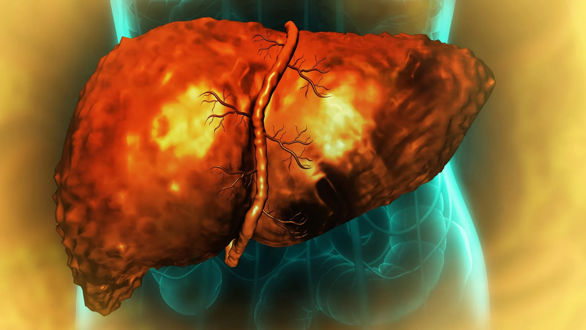
Excessive alcohol consumption inflicts profound damage on the liver, one of the body’s most resilient organs, by critically impairing its remarkable ability to repair and rebuild itself. New groundbreaking research reveals that alcohol-induced injury can trap liver cells in a persistent, abnormal intermediate state, rendering them incapable of normal function or completing the vital regeneration process, even after an individual ceases drinking. This cellular "limbo" appears to be orchestrated by chronic inflammation that disrupts RNA splicing, a fundamental cellular process essential for translating genetic blueprints into functional proteins. The implications of these findings, published in the esteemed journal Nature Communications, are far-reaching, potentially paving the way for novel diagnostic tools and therapeutic interventions for severe alcohol-associated liver disease (ALD), a leading global health crisis.
The Liver’s Unparalleled Regenerative Capacity Under Siege
The liver stands out among human organs for its extraordinary capacity to regenerate. Following injury, disease, or even surgical removal of a significant portion, the remaining liver cells possess the ability to temporarily alter their identity, proliferate, and then mature back into fully functional cells, restoring the organ’s mass and function. This inherent resilience is crucial for maintaining the liver’s myriad vital roles, including detoxification, metabolism, and protein synthesis.
However, this remarkable regenerative capacity falters dramatically in the context of alcohol-associated liver disease. ALD encompasses a spectrum of liver conditions, from fatty liver disease and alcoholic hepatitis to the more severe and often irreversible cirrhosis. Globally, ALD is the primary driver of liver-related mortality, contributing to an estimated three million deaths annually. The progressive scarring and functional decline characteristic of cirrhosis often necessitate liver transplantation, a procedure fraught with its own challenges, including donor organ scarcity and lifelong immunosuppression.
"For years, clinicians and researchers have observed that the liver’s ability to regenerate is severely compromised in patients suffering from alcoholic hepatitis and cirrhosis," stated Professor Auinash Kalsotra, a distinguished biochemist at the University of Illinois Urbana-Champaign and a co-lead investigator of the study. "Even when patients abstain from alcohol, their livers often fail to recover. The critical missing piece of the puzzle was understanding why this regeneration process halts. Our findings offer a significant leap forward in elucidating these underlying mechanisms, potentially opening doors for interventions before the liver reaches a state of irreversible failure."
Professor Anna Mae Diehl of Duke University School of Medicine, who co-led the study with Professor Kalsotra, echoed this sentiment, emphasizing the long-standing clinical need for a deeper understanding of ALD pathogenesis. "The only definitive treatment for end-stage liver disease is transplantation. If we can pinpoint the molecular reasons behind the liver’s failure to regenerate, we can begin to explore targeted therapeutic strategies to restore its natural healing capabilities."
Unraveling the Cellular Stalemate: Cells Trapped in a Regenerative Limbo
The research team embarked on a meticulous comparative analysis, examining healthy liver tissue samples alongside those obtained from individuals diagnosed with alcohol-associated hepatitis or cirrhosis. These diseased liver samples were generously provided by Johns Hopkins University Hospital, facilitated by an initiative supported by the National Institute on Alcohol Abuse and Alcoholism (NIAAA), a division of the National Institutes of Health (NIH).
A striking and consistent pattern emerged from this comparative analysis. In the diseased liver tissues, cells that had initiated the transition from a mature, functional state towards a more primitive progenitor-like state—a necessary step for regeneration—were found to be unable to complete this crucial transformation. Instead, these cells became ensnared in an intermediate, quasi-progenitor state, a cellular "limbo" from which they could neither function as mature liver cells nor effectively proliferate as true progenitor cells.
"These cells are essentially caught in a developmental deadlock," explained Ullas Chembazhi and Sushant Bangru, graduate students at the University of Illinois and co-first authors of the study. "They are neither fully functional adult cells nor capable of the robust division characteristic of progenitor cells. Because they are not performing their designated tasks, an immense burden is placed on the remaining healthy cells. These healthy cells then attempt to regenerate, but in doing so, they too risk becoming trapped in this unproductive quasi-progenitor state, ultimately leading to organ failure."
This entrapment creates a vicious cycle of cellular dysfunction and organ decline. As more cells succumb to this state of suspended animation, the pool of actively functioning liver cells diminishes, exacerbating the workload on the survivors. This increased demand for function further pressures the remaining cells to regenerate, inadvertently pushing them towards the same regenerative limbo.
The Critical Role of RNA Splicing and the Downfall of ESRP2
To decipher the molecular mechanisms underlying this regenerative blockade, the researchers delved into the intricate processes of protein synthesis and RNA processing within the liver cells. RNA molecules serve as essential intermediaries, carrying genetic instructions from DNA in the cell’s nucleus to the protein-building machinery located in the cytoplasm. A pivotal step in this process is RNA splicing, where non-coding regions (introns) are removed from the RNA transcript, and the coding regions (exons) are precisely joined together. This precise editing dictates the final protein product, influencing its structure, function, and cellular localization.
Unlike many previous studies that focused on overall RNA and protein levels, Professor Kalsotra’s team employed sophisticated deep RNA sequencing and advanced computational analysis to meticulously examine the patterns of RNA splicing. Their investigation revealed a widespread disruption in RNA splicing across thousands of genes in liver cells affected by alcohol-related disease.
"Our analysis demonstrated that RNA was being ‘misspliced’ on a massive scale in alcohol-damaged livers," Professor Kalsotra elaborated. "This widespread missplicing was not confined to a few genes but affected thousands, profoundly impacting the functional capabilities of the proteins produced."
A key culprit identified in this widespread RNA splicing dysfunction was the significant reduction in the levels of a crucial protein known as ESRP2 (Epithelial Splicing Regulatory Protein 2). ESRP2 plays a vital role in ensuring the accurate splicing of RNA molecules. In alcohol-damaged liver cells, the researchers observed a marked deficiency in ESRP2.
The consequences of ESRP2 deficiency were profound and extended beyond simply affecting protein production. The altered RNA splicing patterns, driven by low ESRP2, frequently disrupted the molecular signals that direct proteins to their correct cellular compartments. Proteins must be in the right place within the cell to perform their specific functions.
"Proteins are designed to operate in precise locations within the cell, and this localization is dictated by specific sequences within the protein itself," Professor Kalsotra explained. "We discovered that in many instances, the sequences responsible for directing a protein’s location were being misspliced. This meant that even if the same amount of RNA and protein was present, the protein was not at its intended site of action. For example, crucial proteins required for effective liver regeneration were found to be inappropriately sequestered in the cytoplasm, when their function demanded their presence within the nucleus, where the cell’s DNA resides and gene regulation occurs."
Experimental Validation and the Inflammatory Cascade
To definitively establish the causal link between ESRP2 deficiency and regenerative failure, the researchers conducted experiments using mice genetically engineered to lack the gene responsible for producing ESRP2. These mice exhibited liver injury patterns and a marked inability to regenerate that closely mirrored the pathological observations in human patients with advanced alcohol-related hepatitis.
This experimental evidence strongly implicated reduced ESRP2 levels as a significant contributor to the regenerative block. The subsequent critical question became: what triggers the initial decline in ESRP2 levels? The research team traced the problem back to the pervasive inflammatory response ignited by chronic alcohol consumption.
When the liver metabolizes alcohol, it can trigger direct tissue damage and initiate an inflammatory cascade, attracting immune cells and specialized liver support cells. The study found that these activated cells release a cocktail of inflammatory and growth factors. These signaling molecules, in turn, were found to suppress both the production and the activity of ESRP2 within liver cells.
Therapeutic Horizons and Diagnostic Potential
The discovery of this inflammatory pathway leading to ESRP2 deficiency and subsequent RNA splicing defects opened a promising avenue for potential therapeutic interventions. In laboratory cultures of liver cells, the researchers demonstrated that by using a molecule designed to block the receptor for a key inflammation-promoting factor, they could restore ESRP2 levels and normalize RNA splicing patterns.
This groundbreaking finding suggests that future therapies might not need to directly replace damaged liver tissue. Instead, they could focus on interrupting the inflammatory signals that cripple the liver’s innate regenerative machinery. By dampening the inflammatory response, it may be possible to enable the liver cells to complete their repair processes effectively.
Furthermore, the identification of abnormally spliced RNA molecules holds significant potential for diagnostic applications. These misspliced RNA transcripts could serve as sensitive biomarkers, allowing for the early detection, diagnosis, and monitoring of alcohol-associated liver disease. This could enable timely interventions before irreversible damage occurs.
"We are incredibly hopeful that these findings will serve as a catalyst for future clinical studies," Professor Kalsotra stated. "The ability to utilize these mis-spliced RNAs as diagnostic markers or to develop novel treatments that effectively curb inflammation represents a significant advancement. If we can successfully correct these splicing defects, we may be able to dramatically improve recovery rates and restore function to damaged livers."
The research was a collaborative effort involving a multidisciplinary team of scientists from the University of Illinois Urbana-Champaign, Duke University, and the Chan Zuckerberg Biohub Chicago. Significant contributions were made by graduate students Diptatanu Das and Subhashis Natua, undergraduate students Katelyn Toohill, Ishita Purwar, and Anuprova Bhowmik from the University of Illinois; Brandon Peiffer and Zhaoli Sun from Johns Hopkins University School of Medicine; Aurelia Leona and Yogesh Goyal from Northwestern University; and Rajesh Dutta from Duke University School of Medicine. Funding for this critical research was provided by the National Institutes of Health, the Chan-Zuckerberg Biohub Chicago, the Duke Endowment, and the Muscular Dystrophy Association, underscoring the broad scientific and philanthropic interest in tackling this pervasive health issue.