


Researchers from Denmark and the United Kingdom have unveiled a groundbreaking brain implant, the microfluidic Axialtrode (mAxialtrode), poised to revolutionize neuroscience research and potentially pave the way for novel treatments for neurological disorders like epilepsy. This innovative device, developed through a collaborative effort involving the Technical University of Denmark (DTU), the University of Copenhagen, and University College London (UCL), offers scientists an unprecedented level of precision in studying the intricate workings of the brain. The findings detailing this advancement were recently published in the esteemed journal Advanced Science.
The mAxialtrode distinguishes itself from existing brain implants through its unique design: a long, needle-thin electrode equipped with microscopic channels. This ingenious configuration allows for multiple functional points along the entire length of the implant, enabling researchers to simultaneously record neural activity and deliver therapeutic agents to highly specific locations within the brain. This multi-functional capability addresses a significant limitation of current technologies, which often require multiple separate implants to achieve similar granular control.
A Multifaceted Tool for Unraveling Brain Complexity
Initially envisioned as a sophisticated research instrument, the mAxialtrode holds immense promise for dissecting complex neural processes. Scientists can now investigate the nuanced pathways of signal transmission across different brain layers during critical functions such as memory formation, decision-making, and the complex cascade of events that characterize epileptic seizures. By providing access to multiple neural strata with a single implant, the device facilitates more comprehensive and accurate studies, reducing the inherent variability associated with multiple insertion points.
The implications for understanding conditions like epilepsy are particularly profound. Epilepsy is characterized by abnormal electrical activity in the brain, often involving widespread neuronal networks. The ability to monitor and modulate activity at multiple depths simultaneously could offer invaluable insights into the initiation and propagation of seizures, potentially leading to the identification of new therapeutic targets.
Beyond its research applications, the mAxialtrode is also being eyed for its long-term therapeutic potential. The device’s capacity to deliver medication to precise locations, coupled with its ability to perform electrical or light stimulation, opens up exciting avenues for targeted interventions. For instance, in conditions where localized inflammation or overactivity contributes to symptoms, the mAxialtrode could deliver anti-inflammatory drugs or neuromodulatory agents directly to the affected areas while simultaneously applying localized electrical stimulation to quell aberrant neural firing.
Addressing the Limitations of Conventional Implants
Dr. Kunyang Sui, a postdoctoral researcher who co-developed the mAxialtrode concept with Associate Professor Christos Markos, highlighted the device’s integrated functionality as a key advantage. "Most current brain implants are based on hard materials such as silicon, which can irritate the brain and trigger inflammatory reactions in the tissue," Dr. Sui explained. "The new implant differs in that it is made of soft, plastic-like optical fibers and has a specially angled tip that makes it smaller and reduces the damage caused when it is placed in the brain."
This emphasis on biocompatibility and minimal invasiveness is crucial. Traditional brain implants, often constructed from rigid materials like silicon or metal, can cause chronic inflammation and scarring as the brain tissue reacts to the foreign body. Over time, these reactions can compromise the implant’s function and lead to adverse side effects for the patient. The mAxialtrode’s flexible, plastic-like composition is designed to move with the brain tissue, significantly reducing mechanical stress and the potential for inflammatory responses.
The development of the mAxialtrode represents a significant leap forward from conventional optical fibers, which have been a mainstay in brain research for years. These standard fibers, typically made of glass or plastic, are used to deliver light deep into the brain for optogenetic stimulation – a technique that uses light to control genetically modified neurons. However, their primary limitation lies in their unidirectional interaction with brain tissue.
"Conventional fibers, however, have an important limitation. They typically interact with brain tissue only at the end of the fiber, meaning researchers can stimulate or monitor just one location at a time," Dr. Sui elaborated. "The outermost end is called the distal tip, or the ‘nose’ of the fiber. Light is emitted and contact with brain tissue occurs only at this point. As a result, scientists may be limited to measuring or stimulating one brain layer at a time, even though many brain functions depend on communication among several layers and deeper structures." This singular point of interaction restricts the ability to study the complex, distributed nature of neural networks.
The Ingenious Design of the mAxialtrode
The creation of the mAxialtrode is a testament to advanced microfabrication techniques. The process begins with a larger polymer rod, which is then heated and meticulously drawn into an extremely thin fiber. This process, akin to creating a delicate sugar strand but with unparalleled precision, results in a fiber measuring less than half a millimeter in diameter.
At the heart of this miniature marvel is a light-conducting core, designed to transmit light for stimulation purposes. Crucially, this core is enveloped by eight microscopic channels. These channels are not merely passive conduits; they are engineered to transport liquids, enabling the precise delivery of drugs or other therapeutic agents. Furthermore, these channels can house incredibly thin metal wires, allowing for the simultaneous recording of electrical activity from the brain.
This integrated design is what sets the mAxialtrode apart. It consolidates multiple research and therapeutic modalities – light delivery, fluid infusion, and electrical recording – into a single, slender implant. This not only simplifies experimental setups and surgical procedures but also minimizes the physical footprint within the delicate brain environment. The flexibility of the fiber is also a critical design element, allowing it to adapt to the natural movements of brain tissue, thereby further mitigating the risk of mechanical irritation and inflammation.
Rigorous Testing in a Living Model
To validate its efficacy and safety, the mAxialtrode underwent extensive testing, including "in vivo" experiments in living mice. In these studies, the electrode was surgically implanted into the animals’ brains and connected to various systems, including light sources for stimulation, recording equipment to capture neural signals, and miniature pumps for fluid delivery.
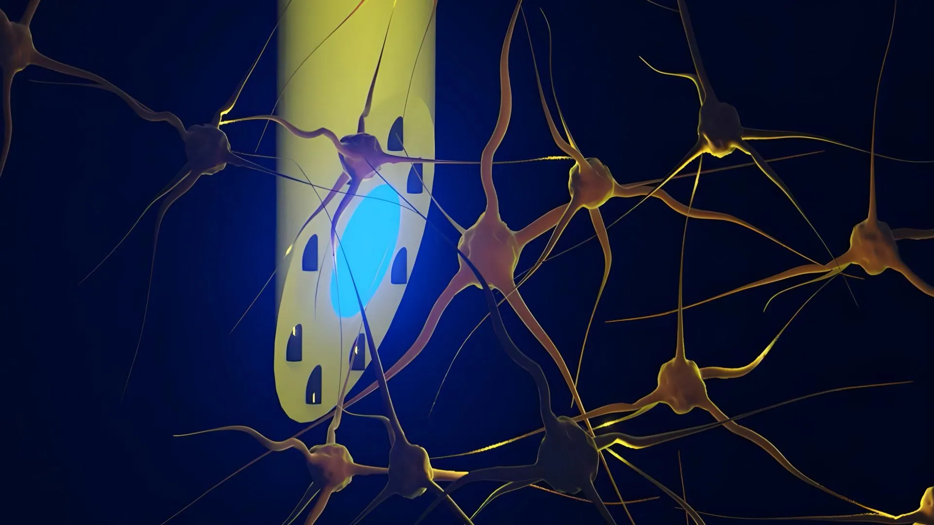
The results of these experiments were highly encouraging. The mAxialtrode successfully stimulated nerve cells using both blue and red light, demonstrating its versatility in optogenetic applications. Simultaneously, researchers were able to record electrical activity from distinct brain regions, encompassing both superficial layers like the cerebral cortex and deeper structures such as the hippocampus. This ability to monitor activity across different anatomical locations with a single device is a significant advancement.
Perhaps most strikingly, the mAxialtrode demonstrated its capacity for precise localized drug delivery. Researchers were able to inject different substances at separate depths, with delivery points spaced approximately three millimeters apart. All of these complex operations – stimulation, recording, and infusion – were performed using the single, lightweight fiber. The mice tolerated the implant well, showing no discernible signs of discomfort, which is a crucial indicator of the device’s biocompatibility.
Illuminating Pathways to Neurological Treatments
The "in vivo" experiments and neurophysiological validation were conducted in close collaboration with Associate Professor Rune W. Berg from the University of Copenhagen and Associate Professor Rob C. Wykes from University College London. Their expertise in analyzing neural circuits and developing models relevant to epilepsy was instrumental in demonstrating the mAxialtrode’s potential in this specific neurological condition.
The collaborative nature of this research underscores the interdisciplinary effort required to push the boundaries of neuroscience. By pooling knowledge in microfluidics, materials science, electrical engineering, and neurobiology, the team has created a technology with far-reaching implications.
The research team is now actively pursuing patent protection for the underlying technology of the brain electrode. Concurrently, they are initiating the process of exploring the regulatory pathways and requirements necessary for eventual clinical translation. While Dr. Sui cautions that the technology is still far from routine clinical use, emphasizing the need for extensive testing, further development, and stringent regulatory approvals, the initial findings represent a significant stride towards that goal.
The potential applications extend beyond epilepsy. The ability to precisely target neural circuits could benefit a wide range of neurological and psychiatric conditions, including Parkinson’s disease, chronic pain, depression, and anxiety disorders. By offering a more refined way to study the brain, the mAxialtrode could unlock fundamental understandings of neural function and dysfunction, leading to the development of more effective and personalized treatments. The journey from laboratory innovation to clinical application is often lengthy and complex, but the mAxialtrode has opened a promising new chapter in the quest to understand and heal the human brain.