


Scientists at Johns Hopkins Medicine have uncovered compelling new evidence suggesting that miniature brain tissues, cultivated from the cells of individuals with Alzheimer’s disease, hold significant potential in predicting how specific patients might respond to medications designed to manage the condition’s challenging neuropsychiatric symptoms. This pioneering research, which utilized patient-derived brain organoids, not only deepens our understanding of Alzheimer’s but also paves the way for more precise and individualized treatment strategies for the millions affected by this devastating neurodegenerative disorder. The findings were recently published in the esteemed journal Alzheimer’s & Dementia: The Journal of the Alzheimer’s Association, with partial funding provided by the National Institutes of Health (NIH).
The Promise of Miniature Brain Models for Precision Medicine
Alzheimer’s disease, the most prevalent form of dementia, currently affects over 7 million Americans, a number projected to rise significantly in the coming decades as the population ages. While a cure remains elusive, managing the often-debilitating neuropsychiatric symptoms—such as anxiety, depression, agitation, and hallucinations—is crucial for improving the quality of life for patients and their caregivers. These symptoms are nearly universal among individuals with Alzheimer’s, yet their response to standard pharmacological interventions, like selective serotonin reuptake inhibitors (SSRIs), can be highly variable. This variability underscores a critical unmet need for personalized treatment approaches.
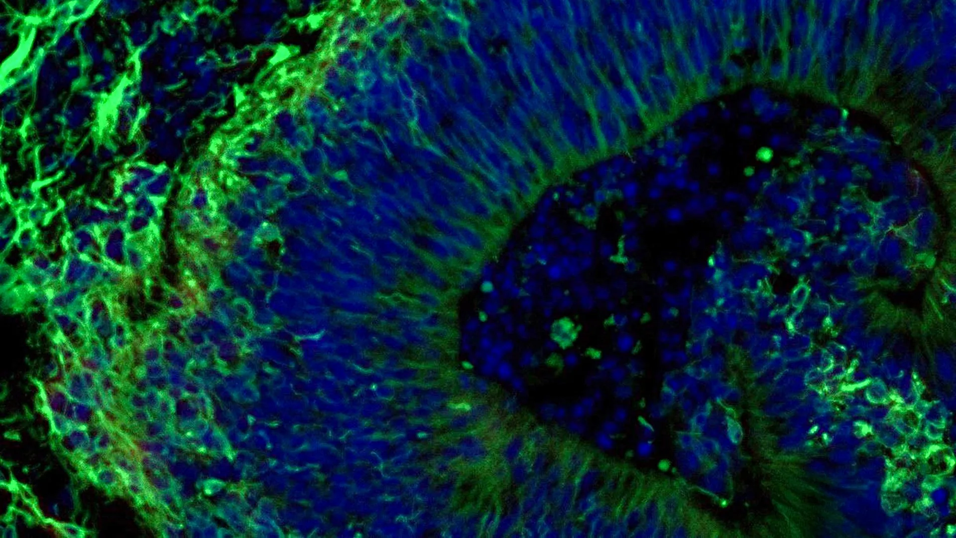
The Johns Hopkins team’s investigation centered on laboratory-grown brain tissues, known as organoids, which are essentially three-dimensional, self-organizing structures that mimic aspects of the human brain. By using organoids derived from individuals with Alzheimer’s, researchers can create controlled environments to study disease mechanisms and test potential therapeutic interventions in ways not possible with traditional cell cultures or animal models. This approach offers a unique window into the complex cellular and molecular changes that characterize Alzheimer’s disease.
"Our study suggests that large-scale, patient-derived brain organoids and the vesicles they secrete can help us stage Alzheimer’s disease, investigate the mechanisms that drive it and assess how patient subgroups may respond to different treatments," stated Dr. Vasiliki Machairaki, the study’s lead investigator and an associate professor of genetic medicine at Johns Hopkins University School of Medicine. "This represents a significant step toward developing more precise, targeted therapies."
Replicating Brain Function in a Dish: The Organoid Creation Process
The research focused on hindbrain organoids, a region of the brain at the back of the skull responsible for regulating fundamental life-sustaining functions like breathing, sleep cycles, and heart rate. The scientists aimed to determine if these miniature brain models could reveal molecular signatures indicating whether a common SSRI, escitalopram oxalate, might effectively alleviate Alzheimer’s-related neuropsychiatric symptoms.
The genesis of these organoids began with blood samples meticulously collected from individuals diagnosed with Alzheimer’s disease, with their informed consent, at the NIH-funded Johns Hopkins Alzheimer’s Disease Research Center. These blood samples were then subjected to a sophisticated reprogramming process. Blood cells were induced to revert to a pluripotent stem cell-like state, transforming them into induced pluripotent stem cells (iPSCs). These iPSCs possess the remarkable ability to differentiate into virtually any cell type in the body, including the specialized neurons that form the intricate network of the brain.
Utilizing iPSCs derived from both Alzheimer’s patients and healthy control individuals, the researchers cultivated hindbrain organoids. These organoids were carefully guided to self-organize into small, pea-sized clusters of brain tissue, remarkably resembling the anatomical structure of the hindbrain. The study’s scale was notable, encompassing hundreds of organoids, each representing an individual patient with Alzheimer’s or a healthy participant. Dr. Machairaki highlighted that this likely represents one of the most extensive brain organoid studies conducted to date in the field of Alzheimer’s research, providing a robust dataset for analysis.
Unveiling Molecular Hallmarks of Alzheimer’s in Organoids
A key finding of the study was that the patient-derived organoids effectively recapitulated several critical biological characteristics of Alzheimer’s disease at the molecular level. When compared to organoids generated from healthy individuals, those grown from the cells of people with Alzheimer’s exhibited distinct differences in the expression and function of proteins involved in crucial brain processes. These included proteins associated with neuronal communication, neuroinflammation—a known contributor to Alzheimer’s pathology—and other pathways implicated in the disease’s progression.
Following the characterization of these disease-specific molecular signatures, the researchers proceeded to treat a subset of the organoids with escitalopram oxalate, a widely prescribed SSRI antidepressant. The results were illuminating. In some organoids derived from Alzheimer’s patients, the medication appeared to stimulate the production of proteins integral to serotonin signaling and intercellular communication. These are precisely the pathways that SSRIs are designed to modulate. However, a significant observation was that other organoids derived from Alzheimer’s patients showed little to no discernible molecular response to the drug.
"We used these organoids to model how some patients’ tissue may respond to a commonly prescribed SSRI," Dr. Machairaki explained. "On a large-scale level, our model may eventually be used to identify subgroups of patients, based on underlying molecular mechanisms, who are more likely to respond to certain drugs and thus help us to create precise, targeted treatments in the long run." This suggests that the organoids can serve as a personalized platform to predict drug efficacy, potentially averting ineffective treatments and associated side effects for patients.
Extracellular Vesicles: Tiny Messengers with Diagnostic Potential
Beyond cellular responses, the Johns Hopkins team also explored the role of extracellular vesicles (EVs) released by the organoids. EVs are tiny, membrane-bound particles secreted by cells that act as intercellular communicators, carrying a cargo of proteins, RNA, and other molecules. The researchers hypothesized that these EVs might serve as valuable biomarkers for Alzheimer’s disease or offer insights into how brain tissue responds to therapeutic interventions.
Before and after administering escitalopram treatment to the organoids, scientists meticulously analyzed the protein content within the EVs released by both patient-derived and healthy control organoids. The findings revealed that these EVs contained proteins critical for fundamental brain activities, including neuronal communication, memory formation, and the release of neurotransmitters.
Crucially, organoids derived from individuals with Alzheimer’s disease showed marked alterations in several disease-associated proteins within their secreted EVs. Specifically, levels of proteins such as RAB3A, NSF, and ATCAY were found to be diminished in the EVs from Alzheimer’s organoids. These proteins play vital roles in the normal functioning of synaptic signaling, the critical process by which neurons transmit information.
Following escitalopram treatment, the study observed an increase in the levels of certain proteins within the EVs in specific samples. These changes were particularly pronounced in proteins associated with serotonin signaling and synaptic pathways, the very targets of antidepressant medications. The differential response observed across organoids—some exhibiting robust molecular changes in their EVs and others showing minimal to no alteration—further strengthened the hypothesis that EVs could be harnessed to predict drug response.
"This variation raises the possibility that extracellular vesicles from brain organoids could eventually help identify which patients are most likely to benefit from a particular treatment," Dr. Machairaki noted. This opens up a promising avenue for developing non-invasive diagnostic tools.
The Road Ahead: Enhancing Organoid Realism and Clinical Application
The current study represents a significant leap forward, but Dr. Machairaki and her team are already looking toward the future, aiming to develop even more sophisticated and realistic brain organoid models. Future iterations will incorporate additional cell types, such as immune cells (microglia), which are heavily implicated in Alzheimer’s pathology, and vascular-like networks that mimic the intricate structure of blood vessels in the brain. The inclusion of these components is expected to make the organoids even more representative of living human brain tissue, thereby enhancing their predictive power.
With continued research and refinement, Dr. Machairaki envisions that extracellular vesicles derived from these advanced organoids could one day function as a form of "liquid biopsy." Such a non-invasive test could revolutionize Alzheimer’s diagnosis, enabling earlier detection, more accurate staging of disease progression, and the identification of specific disease subtypes, each potentially requiring a tailored therapeutic approach.
This groundbreaking research, supported by substantial funding from the National Institutes of Health, the Paul G. Allen Frontiers Foundation, and the Richman Family Precision Medicine Center of Excellence in Alzheimer’s Disease at Johns Hopkins University, underscores the transformative potential of patient-derived organoid technology. While acknowledging that the current study is an early but vital step, the implications for the future of Alzheimer’s care are profound, offering a beacon of hope for more personalized, effective, and ultimately, more humane management of this complex disease. The collaborative effort involved scientists from Johns Hopkins, Tymora Analytical Operations, and the University of Rochester School of Medicine and Dentistry, highlighting the multidisciplinary nature of modern biomedical research.