


Stanford University scientists have identified a groundbreaking new type of immune cell in planarian flatworms that self-destructs by exploding, a phenomenon they’ve termed "ruptosis." This dramatic cellular demolition occurs with astonishing speed and completeness, eliminating the offending cell and its immediate surroundings within minutes, leaving virtually no trace. The discovery, published in the esteemed journal Cell, offers a tantalizing glimpse into ancient immune strategies and could potentially pave the way for novel therapeutic approaches in modern medicine.
The unique capabilities of planarian flatworms, renowned for their unparalleled regenerative powers, have long fascinated scientists. These seemingly simple aquatic creatures can regrow entire body parts, and even complete organisms, from minuscule fragments. Understanding how their immune systems have evolved and adapted over hundreds of millions of years provides a fertile ground for investigating biological mechanisms that might be lost or suppressed in more complex animals, including humans.
Discovery of Ruptoblasts: A Violent Immune Response
The identification of these explosive cells, officially named "ruptoblasts" by the research team, stemmed from investigations into a fundamental question in flatworm biology: their ability to distinguish self from non-self. Chew Chai, a postdoctoral researcher in the associate professor Bo Wang’s laboratory, was at the forefront of this inquiry.
To probe this immune recognition, Chai meticulously sliced planarian flatworms lengthwise and then fused each half with tissue from a different, unrelated individual. The hypothesis was that if flatworms possessed an immune system capable of recognizing foreign tissue, they would reject the grafted segment. This experiment mirrored the challenges faced in human organ transplantation, where the recipient’s immune system often mounts a formidable defense against donor tissue.
As anticipated, the "Frankenstein" flatworms exhibited a pronounced rejection response to the foreign tissue. However, the cellular mechanisms underlying this rejection were far from conventional. Instead of the more gradual inflammatory responses typically observed in vertebrates, the flatworms displayed a violent and immediate cellular reaction. "It’s this huge inflammatory response. Like there’s a fire and an alarm goes off, and the cells just blow up," described Chai, the lead author of the study.
The Role of Activin in Triggering Ruptosis
Further investigation revealed a crucial trigger for this dramatic cellular self-destruction: the hormone activin. Prior research had already established activin’s pivotal role in flatworm survival, influencing both regeneration and reproduction. The Stanford team observed that as the fused worms began to reject the foreign tissue, there was a significant surge in activin levels. This hormonal spike was followed by a chronic inflammatory state that, while not immediately fatal, ultimately led to the worms’ demise within days.
Crucially, Chai found that injecting activin into healthy, unfused flatworms induced a similar inflammatory cascade, suggesting that activin acts as a direct signal for this aggressive immune response. This finding was pivotal in understanding the conditions under which ruptoblasts are activated.
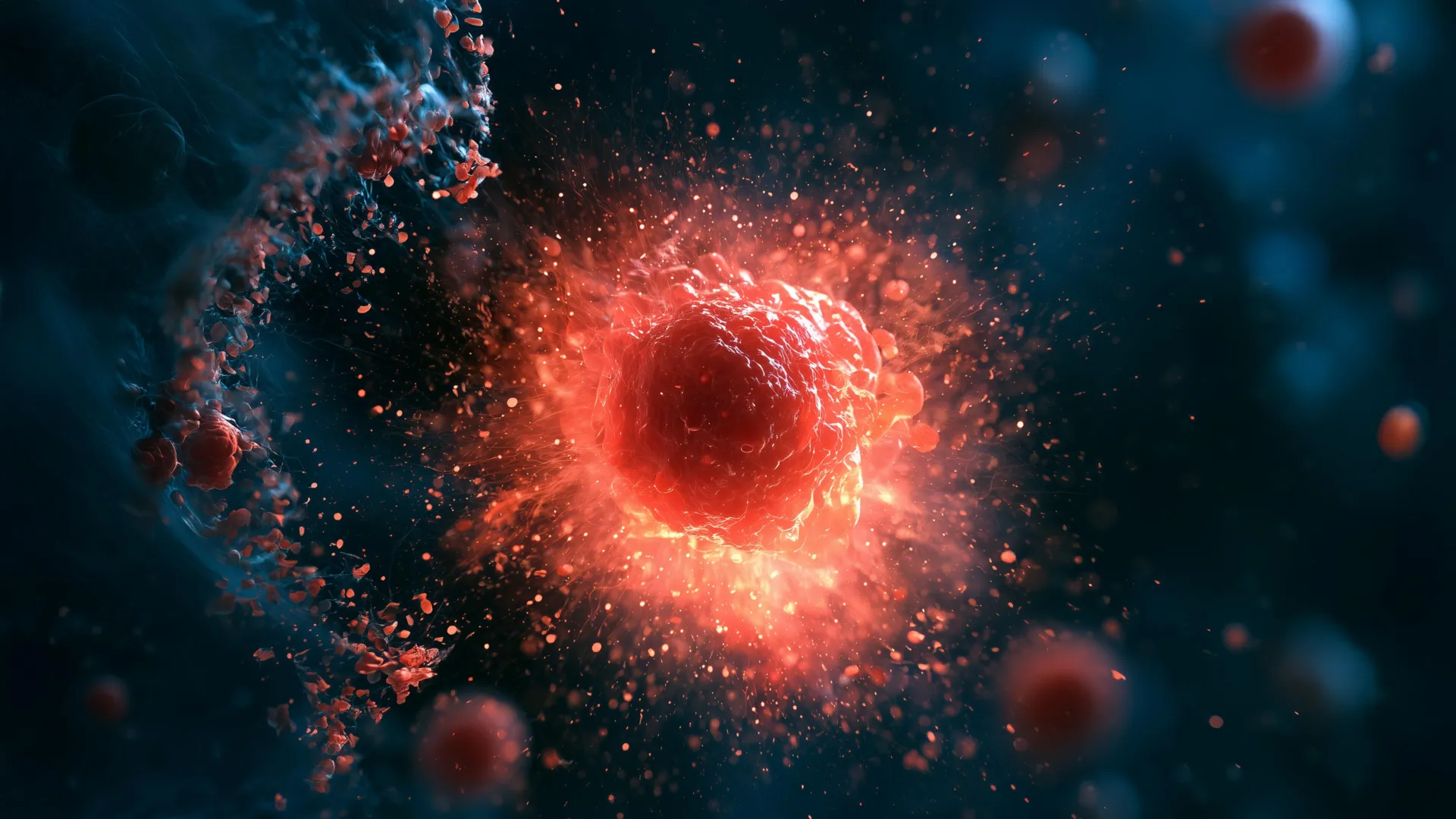
To delve deeper into the cellular mechanisms, Chai employed advanced live-cell microscopy and flow cytometry. By labeling cells with fluorescent dyes, she was able to track and isolate those that responded to the presence of activin. The results were startling: a small population of cells underwent a rapid and violent transformation. They burst open, releasing their contents and effectively destroying neighboring cells before vanishing entirely, a process that took less than five minutes. This rapid, explosive cell death was subsequently termed "ruptosis."
Ruptosis: A Uniquely Rapid Form of Cell Death
The defining characteristic of ruptosis, according to the researchers, is its extraordinary speed and completeness, differentiating it from other known forms of programmed cell death. While some mammalian cells and bacteria can undergo explosive cell death, the timescale is considerably longer, often spanning several hours as harmful substances are slowly leached out. "Ruptosis happens within seconds to minutes," emphasized Chai, highlighting the stark temporal contrast.
This rapid detonation transforms each ruptoblast into a highly localized and potent weapon. Rather than a slow leakage of toxins, the cell unleashes its destructive payload almost instantaneously. This concentrated burst of activity appears to be a highly efficient, albeit destructive, method of eliminating immediate threats.
Ruptoblasts as Multi-Target Weapons
To assess the destructive capacity of ruptoblasts, the researchers subjected them to tests against a variety of targets, including E. coli bacteria, human kidney cells, and mouse blood cells. In each instance, the ruptoblasts proved highly effective, successfully destroying all tested cell types.
Importantly, the damage inflicted by ruptoblasts remained confined to cells in close proximity to the explosion. The destructive event did not propagate through a chain reaction, nor did it leave behind persistent toxicity in the surrounding environment. This capacity for a powerful, yet tightly contained, attack suggests potential applications in future medical treatments. For instance, such a mechanism could be harnessed to target localized bacterial infections or malignant tumors, minimizing collateral damage to healthy tissues.
Ancient Origins and Evolutionary Significance
The research also revealed significant differences between ruptoblasts and more familiar immune cells. Unlike hematopoietic cells such as T cells and neutrophils, which originate from bone marrow, ruptoblasts are classified as glandular cells. This distinction points to a fundamentally different evolutionary origin and functional pathway.
The researchers hypothesize that ruptoblasts represent an ancient immune strategy, originating early in animal evolution. Their search for similar cells in other species revealed their presence primarily in basal bilaterians, such as flatworms, suggesting they may have been lost or superseded in more derived lineages.
A compelling theory proposed by Chai suggests that vertebrates may have evolved away from this strategy due to the inherent difficulty in repairing the extensive damage caused by ruptosis. In contrast, flatworms, with their abundant stem cells and remarkable regenerative capabilities, are exceptionally well-equipped to swiftly replace damaged tissues. This evolutionary divergence highlights the diverse array of immune mechanisms that have arisen in response to varying environmental pressures and biological constraints.
"It demonstrates there’s lots of different immune mechanisms out there. There’s all these animals that live in an environment where there’s lots of bacteria, lots of viruses, and we know so little about their immune mechanisms," stated Bo Wang. The findings underscore the vast untapped potential of studying organisms that are not traditionally considered model systems in scientific research.
Broader Implications for Medicine and Research
The discovery of ruptoblasts and their explosive mechanism of action opens up new avenues for understanding the complexities of the immune system. It challenges conventional notions of cellular defense and suggests that nature has devised remarkably potent and rapid methods for combating threats.
The ability of ruptoblasts to deliver a swift and localized destructive blow could inspire novel therapeutic strategies. Imagine engineered cells that could be deployed to precisely eliminate cancerous cells or antibiotic-resistant bacteria, mirroring the targeted action of ruptoblasts but with controlled efficacy and safety.
Furthermore, the research emphasizes the importance of exploring the biological intricacies of diverse organisms. The seemingly simple planarian flatworm, through its unique regenerative and immune capabilities, has provided profound insights into fundamental biological processes. This broader perspective is crucial for addressing some of medicine’s most intractable challenges. As Wang noted, "investigating a wider range of organisms may inspire new approaches to some of medicine’s most challenging problems."
Acknowledgements and Funding
The groundbreaking research was a collaborative effort involving numerous scientists. Additional co-authors from Stanford University include postdoctoral scholar Souradeep Sarkar; former Undergraduate Visiting Research Program scholar Lihan Zhong; Dania Nanes Sarfati, PhD ’24; Christine Jacobs-Wagner, the Dennis Cunningham Professor and professor of biology in the Schools of Engineering and Medicine and of microbiology and immunology in the School of Medicine; and Hawa Racine Thiam, assistant professor of bioengineering in the Schools of Engineering and Medicine and of microbiology and immunology in the School of Medicine. Significant contributions also came from Benyamin Rosental and other colleagues at Ben Gurion University of the Negev, who served as co-senior author.
Institutional affiliations highlight the interdisciplinary nature of the work, with several researchers holding appointments within Stanford Bio-X, the Wu Tsai Neurosciences Institute, and the Sarafan ChEM-H institute.
The research was generously supported by a range of prestigious funding bodies, including a National Science Foundation Graduate Research Fellowship, a Stanford Graduate Fellowship, a Stanford DARE fellowship, a Human Frontier Science Program grant, a National Institutes of Health grant, and the European Research Council. This collective support underscores the significance and potential impact of this discovery within the scientific community.