


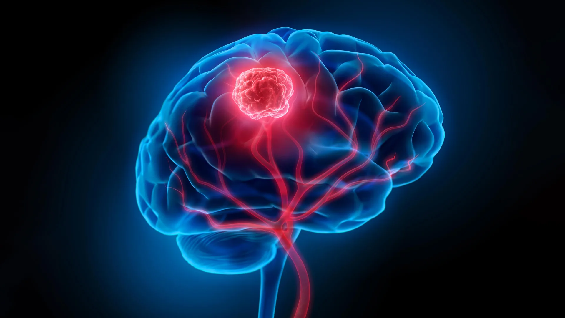
Glioblastoma, the most aggressive form of brain cancer, presents a formidable challenge to medical science due to its invasive nature and the protective barriers of the brain. For decades, the battle against this devastating disease has been hampered by the inability to completely remove cancerous growths without causing irreparable damage to healthy brain tissue. Furthermore, the notorious blood-brain barrier, a biological shield designed to protect the brain, significantly impedes the delivery of therapeutic agents, including chemotherapy drugs and radiation. These combined obstacles contribute to a grim five-year survival rate, hovering at a mere 7 percent, a stark statistic underscoring the urgent need for innovative treatment strategies.
However, a groundbreaking development from a collaborative effort between researchers at the University of Technology Sydney (UTS), Harvard University, and Henan University is poised to redefine the landscape of glioblastoma treatment. Published in the esteemed journal Science Translational Medicine, their work introduces a novel "double-punch" nanozyme platform, a sophisticated system of smart nanoparticles designed to simultaneously address the critical challenges of surgical precision and post-operative eradication of residual cancer cells. This innovative approach, meticulously engineered, offers a dual-action capability within a single, integrated system.
A Sequential Symphony of Nanotechnology
At the heart of this revolutionary platform lies an exceptionally thin, two-dimensional sheet composed of individual atoms precisely arranged using advanced semiconductor manufacturing techniques. This atomic-level engineering imbues the material with a remarkable ability to transition between two distinct, yet complementary, functions. Initially, it serves as an ultra-sensitive imaging agent during surgical procedures, guiding surgeons with unprecedented accuracy. Following the removal of visible tumor masses, the same nanoparticle system is repurposed to deliver a targeted therapeutic intervention, eliminating any microscopic cancer cells that may have escaped surgical detection. Crucially, both of these powerful functions are activated by the same wavelength of near-infrared light, simplifying the treatment protocol and enhancing its efficacy.
Dr. Bingyang Shi, Chair Professor of Nanomedicine at UTS, elaborated on the ingenuity of the design, stating, "We’ve engineered a single material that does two jobs in sequence. It’s a precise guide for the surgeon during the operation, and then a targeted clean-up treatment afterward." This sequential functionality represents a significant leap forward, moving beyond single-purpose treatments to a more comprehensive, multi-faceted attack against the insidious nature of glioblastoma.
Illuminating the Unseen: Enhanced Surgical Visualization
The initial phase of the treatment leverages the nanoparticle’s advanced imaging capabilities. "During surgery, it functions as a highly sensitive imaging agent," explained Professor Shi. A specially engineered fluorescent dye integrated into the nanoparticle sheet emits a distinct glow when exposed to near-infrared light, a spectrum invisible to the human eye. This fluorescence allows surgeons to visualize individual tumor cell clusters with remarkable clarity, even those as small as 44 micrometers. This level of resolution surpasses the capabilities of current clinical imaging tools, enabling surgeons to differentiate cancerous tissue from healthy brain matter with unparalleled precision.
Moreover, the nanoparticle platform is further enhanced by a targeting molecule. This crucial component facilitates the crossing of the formidable blood-brain barrier, a feat that significantly enhances drug delivery in conventional treatments. Once across, this molecule directs the nanoparticles to selectively accumulate within glioma cells, ensuring that the imaging signal is concentrated precisely where it is needed most. This targeted accumulation not only improves the accuracy of visualization but also minimizes potential off-target effects, a critical consideration in delicate neurosurgical procedures. The ability to "see" these microscopic clusters before they become clinically significant is a game-changer, potentially reducing the likelihood of incomplete tumor resection.
Post-Operative Annihilation: A Targeted Therapeutic Strike
Once the visible tumor has been meticulously removed, the nanoparticle platform undergoes a transformation in its role. The same material, now residing within the surgical cavity, is reactivated by the familiar near-infrared light. This activation initiates a potent phototherapy, designed to eradicate any remaining microscopic cancer cells. The mechanism is a testament to elegant biochemical engineering: the platinum atoms embedded within the nanoparticles catalyze a reaction with the tumor’s own hydrogen peroxide, generating oxygen. This oxygenation counteracts the typically hypoxic (low-oxygen) environment that often shields glioblastoma cells from conventional treatments, rendering them more vulnerable.
Simultaneously, the near-infrared light triggers the generation of heat and reactive oxygen species, potent agents that are highly effective in destroying microscopic cancer cells. This dual-action phototherapy ensures that even the most elusive cancer remnants are targeted and eliminated, thereby significantly reducing the risk of tumor recurrence. The strategic targeting of these residual cells addresses one of the most persistent and devastating aspects of glioblastoma: the tendency for the cancer to return, often more aggressively, after initial treatment.
A Glimpse into the Future: Preclinical Successes and the Road Ahead
The efficacy of this innovative nanoparticle platform has been rigorously tested in preclinical studies using mouse models of glioblastoma. The results have been profoundly encouraging. In experiments where the nanoparticle treatment was administered following surgery, tumor recurrence was significantly reduced. Remarkably, every mouse in the treated group survived for at least 60 days, a substantial improvement compared to the 42-day survival observed in mice that underwent surgery alone. Furthermore, comprehensive follow-up assessments revealed no detectable neurological or motor impairments associated with the treatment, suggesting a favorable safety profile.
These preclinical findings offer a beacon of hope, demonstrating the potential of this dual-function nanoparticle system to revolutionize glioblastoma treatment. The ability to both visualize microscopic tumors with unprecedented accuracy and then therapeutically eliminate residual cancer cells addresses the fundamental limitations that have plagued glioblastoma therapy for so long.
The Broader Implications and the Path to Clinical Application
The implications of this research extend far beyond the immediate context of glioblastoma. The underlying principles of engineering multi-functional nanoparticles that can navigate biological barriers, target specific cells, and deliver sequential therapeutic actions hold immense promise for a wide range of complex diseases. The development of materials that can seamlessly transition between diagnostic and therapeutic roles represents a paradigm shift in personalized medicine, potentially paving the way for more effective and less invasive treatments for various cancers and other debilitating conditions.
However, the researchers are quick to temper enthusiasm with a clear understanding of the research’s current stage. "The results are very encouraging, but this is still early-stage research carried out in mouse models, not in people – and that distinction is important," Professor Shi emphasized. The transition from animal models to human clinical trials is a complex and lengthy process, involving extensive safety and efficacy testing. The performance of the nanoparticle platform will need to be validated at the scale of a human brain, a significantly more intricate biological environment.
Looking Forward: A Collaborative Endeavor for a Brighter Tomorrow
The collaborative spirit that fueled this innovation is a testament to the power of interdisciplinary research. The integration of expertise from engineering, medicine, and materials science has been critical in developing such a sophisticated and effective solution. As the research progresses, further collaboration will be essential to navigate the regulatory pathways and bring this promising technology to patients.
If this technology continues to prove its mettle through rigorous clinical trials, the vision is clear: surgeons empowered with the ability to see and remove more of the tumor during an operation, followed by a targeted therapeutic intervention that eliminates lingering microscopic cells. This represents a significant stride towards reducing tumor recurrence, a challenge that has historically cast a long shadow over the lives of glioblastoma patients and their families. The journey from laboratory breakthrough to widespread clinical application is often arduous, but the potential to fundamentally alter the prognosis for glioblastoma patients makes this pursuit not just a scientific endeavor, but a profound humanitarian mission. The continued investment in and exploration of such innovative nano-therapeutics could usher in a new era in the fight against one of humanity’s most relentless diseases.