


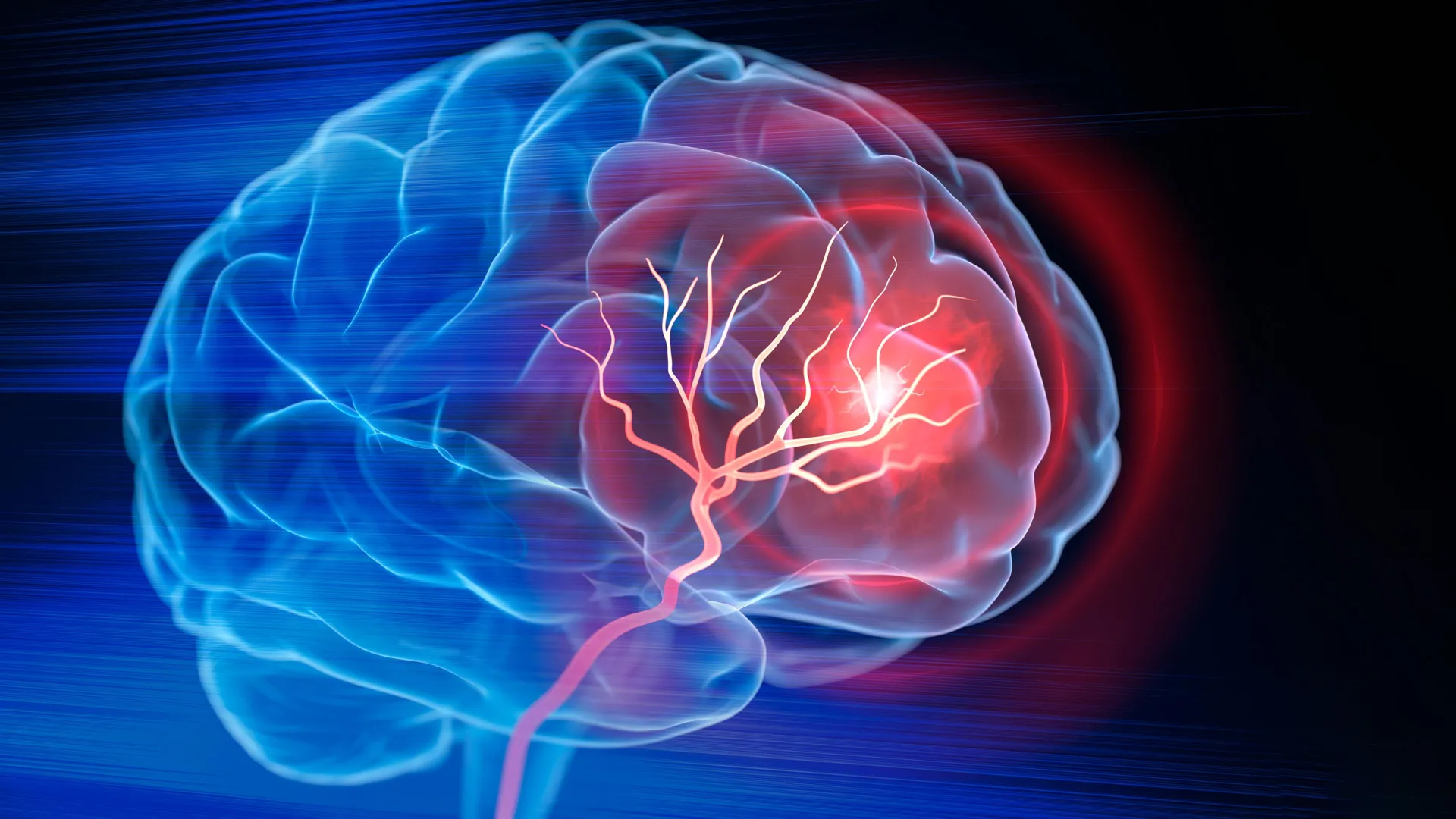
Biomedical engineers at Duke University have achieved a significant breakthrough in stroke treatment, developing an injectable biomaterial designed to transform the post-stroke brain environment and foster recovery. This innovative material, when introduced into the cavity left by lost brain tissue after an ischemic stroke, has demonstrated in preclinical studies the ability to recruit the body’s immune cells, stimulate the formation of new blood vessels, support neural tissue regeneration, and ultimately improve motor function in animal models. The findings, published in the prestigious journal Cell Biomaterials, offer a novel therapeutic strategy for a condition that affects millions globally each year.
The Devastating Impact of Ischemic Stroke
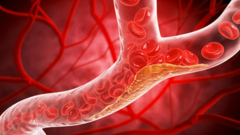
Ischemic strokes represent the most common type of stroke, accounting for approximately 87% of all stroke cases. They occur when a blood clot obstructs blood flow to a region of the brain, depriving brain cells of oxygen and nutrients and leading to cell death. The immediate aftermath of an ischemic stroke is a race against time. Emergency interventions, such as administering clot-dissolving drugs like tPA (tissue plasminogen activator) or performing mechanical thrombectomy procedures to physically remove the clot, are crucial for salvaging viable brain tissue by restoring circulation. However, once brain cells have died, these interventions cannot reverse the loss.
The consequences of significant tissue loss can be profound, leaving behind a physical void within the brain. While rehabilitation plays a vital role in helping surviving neural circuits adapt and compensate for lost function, it does not directly rebuild the damaged or destroyed brain tissue. This leaves a substantial unmet need for therapies that can actively promote the regeneration and repair of the injured brain.
A New Paradigm: Engineering the Injured Space
"Once brain tissue has been lost, restoring blood flow is no longer enough," stated Tatiana Segura, the Robert Plonsey Distinguished Professor of Biomedical Engineering at Duke University and a leading figure in this research. "Our goal is to engineer the injured space so that immune, vascular, and neural repair processes can begin to work together." This statement encapsulates the core philosophy behind the Duke team’s innovation: moving beyond simply addressing the immediate blockage to actively orchestrating a complex, multi-faceted healing response within the brain itself.
The research team’s approach centers on creating a specialized microenvironment within the stroke-induced cavity. This environment is designed to act as a scaffold and a signaling hub, guiding the body’s innate repair mechanisms. By strategically manipulating this space, they aim to overcome the limitations of current stroke treatments, which primarily focus on preventing further damage rather than actively promoting restoration.
The MAPS Scaffold: A Foundation for Healing
The foundation of this novel therapeutic approach is a biomaterial known as microporous annealed particle scaffolds, or MAPS. These scaffolds are constructed from an aggregation of tiny hydrogel microparticles. The unique manufacturing process causes these particles to fuse together, forming a porous, three-dimensional structure. This intricate architecture is crucial, as the interconnected pores provide a physical framework that allows cells to infiltrate, navigate, and colonize the injured area. In essence, the MAPS scaffold acts as a temporary biological support system, providing a stable platform for cellular activity and tissue regeneration.
Building upon earlier successful applications of MAPS in other tissue engineering contexts, the Duke researchers sought to enhance its therapeutic potential by integrating the body’s own immune system into the repair process. They recognized that immune cells, often perceived solely as agents of inflammation, also play critical roles in tissue remodeling and regeneration.
Harnessing the Power of Extracellular Vesicles and Astrocytes
A key component of their strategy involves leveraging the communication network of brain cells, particularly astrocytes. Astrocytes, star-shaped glial cells, are fundamental to normal brain function, providing structural support, regulating the neuronal environment, and playing a crucial role in brain injury response. When the brain is damaged, astrocytes become highly active, communicating with surrounding cells through the release of extracellular vesicles (EVs). These minuscule, membrane-bound sacs are packed with a diverse cargo of proteins, lipids, and genetic material that can profoundly influence the behavior and function of recipient cells.
The Duke team hypothesized that by delivering specific EVs, they could direct the actions of immune cells and other cellular players involved in the healing cascade. Their innovative step was to not merely inject these EVs into the damaged brain but to strategically attach them to the surfaces of the MAPS hydrogel microparticles. This clever immobilization strategy ensures that the therapeutic signals remain concentrated precisely where they are needed – within the scaffold and the stroke cavity. This localized delivery enhances the probability that incoming cells will encounter these crucial signaling molecules, thereby maximizing their impact on the repair process.
"We are not simply placing a material into the brain," Professor Segura emphasized. "We are engineering a local environment that can coordinate several parts of the repair response." This statement underscores the shift from passive implantation to active environmental engineering.
Orchestrating the Immune Response: The Surprising Role of Neutrophils
To guide the immune system effectively, the researchers experimented with various signaling molecules within the EVs, aiming to attract beneficial immune cells, promote blood vessel growth, and enhance overall functional recovery. They identified a particular combination of signaling molecules, Interleukin-4 (IL-4) and C1q, as being exceptionally effective in drawing specific types of immune cells into the injured region.
Among the recruited cells were macrophages, a well-established player in tissue repair. However, the study revealed a surprising and significant role for neutrophils. Neutrophils are typically associated with acute inflammation and tissue damage in the early stages of stroke. Traditionally, their presence has been viewed as detrimental. Yet, the Duke team’s findings suggest a more nuanced and potentially beneficial role for these cells, particularly when present in the engineered microenvironment provided by the MAPS scaffold and influenced by specific signaling cues.
The researchers demonstrated the critical importance of neutrophils by selectively reducing their population. This reduction led to a substantial decline in new blood vessel formation and a less robust remodeling of the scaffold. This observation strongly indicated that neutrophils, under these specific conditions, were actively contributing to the healing process.
Shangjing Xin, the lead scientist of the study and a postdoctoral fellow in Professor Segura’s laboratory, commented on this groundbreaking discovery: "This result changes how we think about neutrophils after stroke. Their role appears to depend on when they arrive, where they are located, and the signals they receive from their surroundings. Our study demonstrates a potential engineering strategy to recruit and retain these cells at the right time." This insight could fundamentally alter our understanding of the inflammatory response following stroke and open new avenues for therapeutic intervention.
Regenerating Vasculature and Neural Connections
The coordinated action of the engineered biomaterial and the recruited immune cells led to remarkable biological changes within the stroke cavity. As immune cells infiltrated the area, the formation of new blood vessels, a process known as angiogenesis, was significantly stimulated throughout the cavity. This increased vascularization is crucial for delivering oxygen and nutrients to the regenerating tissue and clearing waste products.
Furthermore, the researchers observed an increase in axonal fibers, the long projections of nerve cells that transmit signals, both within and surrounding the injured region. This suggests that the biomaterial and the orchestrated cellular activity were not only supporting structural repair but also facilitating the re-establishment of neural connections.
These biological improvements were directly correlated with functional recovery in the treated mice. On a grid-walking test, designed to assess motor coordination and forelimb placement accuracy, mice treated with the optimized MAPS scaffold showed significant improvement. By eight weeks post-treatment, their performance was statistically indistinguishable from that of healthy control mice, and this functional recovery was sustained for the remainder of the study. This demonstrates a potent ability of the engineered biomaterial to promote not just cellular repair but also meaningful functional restoration.
The Indispensable Role of the Scaffold
To ascertain the specific contribution of the MAPS scaffold, the researchers conducted a crucial control experiment. They administered the EVs containing the therapeutic signaling molecules without the presence of the biomaterial. The results were stark: the EVs alone failed to induce comparable levels of blood vessel repair. This finding is critical, as it highlights that the biomaterial is not merely a passive delivery vehicle for the EVs.
Instead, the MAPS scaffold’s porous architecture appears to be essential. Its structure facilitates cell infiltration and colonization, while its ability to concentrate the EV-bound signals within the damaged region creates an optimal microenvironment for repair. The synergistic interaction between the physical scaffold and the localized signaling molecules is therefore fundamental to the observed regenerative effects.
Looking Ahead: Preclinical Promise and Future Directions
While the results are highly promising, the researchers emphasize that this is still an early-stage, preclinical treatment. The current studies have been conducted in mouse models, with the material injected directly into the brain lesion. Significant further research is required before this approach can be considered for human clinical trials.
Key areas for future investigation include comprehensive safety assessments, a deeper understanding of the precise roles and interactions of different immune cell populations in the recovery process, and the evaluation of the treatment’s efficacy in larger animal models that more closely mimic the complexity of human stroke.
Another important aspect for future development is the source of the EVs. Currently, the researchers obtain EVs from lab-grown astrocytes derived from rats. As a next step, Segura’s laboratory is exploring the use of EVs produced by human induced pluripotent stem cell-derived astrocytes. This approach offers the potential for a more scalable and clinically relevant source of therapeutic EVs, while also providing greater control over their genetic and molecular content.
Professor Segura eloquently summarized the overarching vision: "You do not restore an ecosystem simply by containing the initial damage. You have to create the conditions that allow life to return. That is how we think about the stroke cavity. The material is not intended to reproduce the brain itself, but to create an environment where the body’s own cells can enter, communicate, and participate in rebuilding vascularized tissue." This forward-looking perspective emphasizes the potential of this technology to not just patch up damage but to truly rebuild and restore function in the aftermath of a devastating stroke. The successful translation of this research from the lab to the clinic could represent a paradigm shift in how we approach stroke rehabilitation and recovery.