




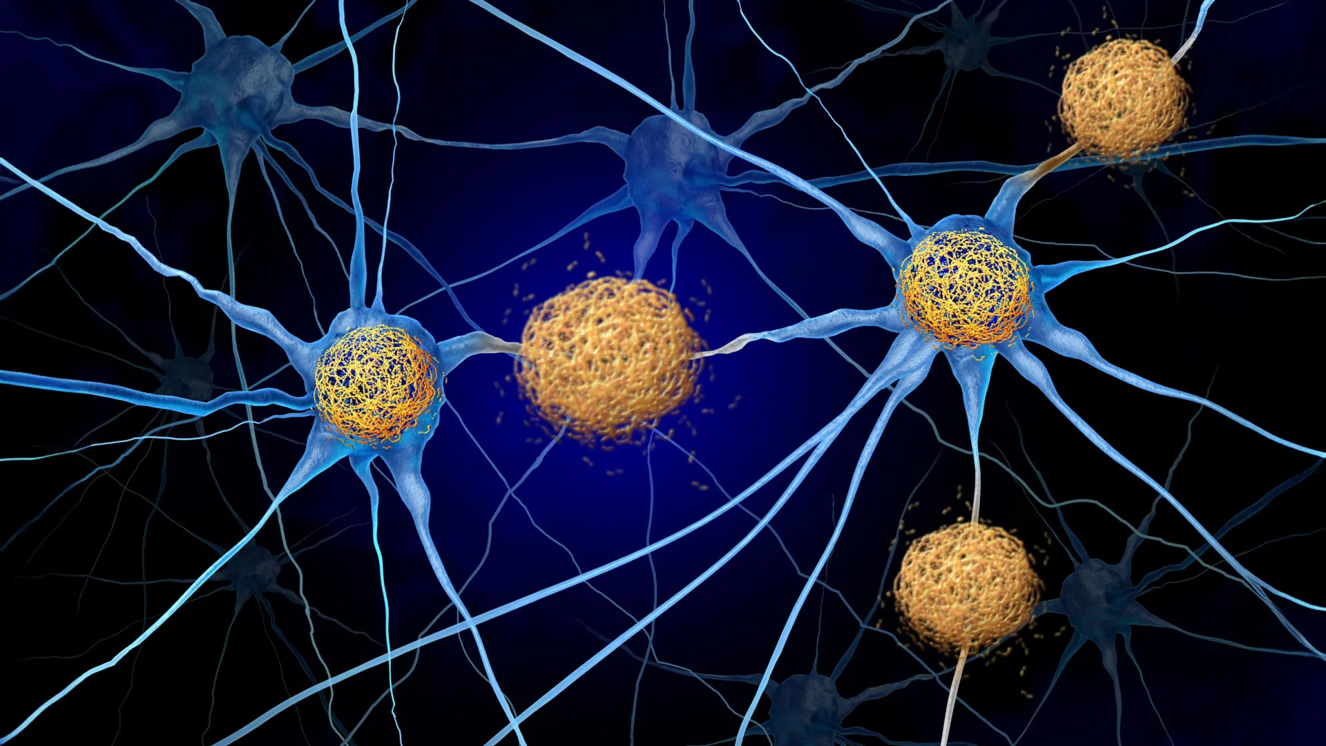
A groundbreaking study, led by scientists from the Department of Psychology at the University of Oslo and published in the prestigious journal Nature Neuroscience, suggests that sophisticated brain imaging techniques could identify changes associated with Alzheimer’s disease significantly earlier than previously understood, potentially up to seven years prior to current detection capabilities. This research challenges the established timeline for Alzheimer’s progression and opens new avenues for early intervention and therapeutic development.
Rethinking the Alzheimer’s Timeline: A Paradigm Shift in Detection
The study’s findings indicate that the current "gold standard" for Alzheimer’s imaging, amyloid-positron emission tomography (PET) scans, which are designed to detect the buildup of amyloid plaques in the brain, may not be sensitive enough to capture the very earliest pathological changes. Researchers have identified distinct structural alterations in the brain that appear at least seven years before amyloid plaques become detectable on these scans. This suggests a more protracted and complex initiation phase for the disease than was previously modeled.
James Michael Roe, the study’s lead researcher during his tenure as a postdoctoral researcher at the Center for Lifespan Changes in Brain and Cognition (LCBC) at the University of Oslo, and now International Scientific Lead at Cercare Medical, emphasized the significance of these discoveries. "We found that structural changes in the brain occur many years before high levels of plaque are seen on PET scans, which is the brain scan currently used to identify the earliest signs of Alzheimer’s disease," Roe stated. This observation implies that the underlying biological processes driving Alzheimer’s may be silently progressing for a considerable period before the characteristic amyloid pathology becomes evident.
Uncovering Subsurface Scars: The Power of Longitudinal MRI
To unravel these subtle, preclinical changes, the research team embarked on an ambitious, long-term observational study. They meticulously followed a cohort of cognitively healthy individuals, subjecting them to regular magnetic resonance imaging (MRI) scans over a period of nearly two decades. This extensive longitudinal dataset was crucial, enabling the scientists to pinpoint the precise timeframe when amyloid plaques first became detectable and to identify which participants in the study would eventually develop them.
Following this, the researchers conducted a retrospective analysis, delving into the MRI scans collected in the decade preceding the first detection of amyloid plaques. By comparing the brain structure changes in individuals who later developed detectable plaques with those who did not, they were able to isolate the specific structural modifications that preceded plaque formation. This comparative approach allowed for the identification of subtle, yet significant, shifts in brain architecture.
Anders Martin Fjell, Professor at the Department of Psychology and head of the LCBC, highlighted the unprecedented nature of their findings. "The most groundbreaking aspect of this study is that we found structural changes in the brain many years before the first signs of plaque buildup, which is considered to be the earliest sign of Alzheimer’s disease," Fjell remarked. He further elaborated on the unique nature of their study population: "These are cognitively well-functioning older individuals. What is unique here is that we have examined changes in brain structure in the years before the first scan revealed plaques." This focus on asymptomatic individuals is critical for understanding the disease’s genesis.
Implications for Early Detection and Intervention Strategies
The discovery of these pre-plaque structural changes has profound implications for the early detection of Alzheimer’s disease. Roe suggested that the earliest signals identified in brain scans could be invaluable for tracking the disease’s progression long before any cognitive symptoms manifest, thereby facilitating earlier diagnosis. "We found the earliest signal detected on brain scans to date, which could be useful for tracking the disease before symptoms emerge and may help with earlier detection," he stated. This advancement could revolutionize how Alzheimer’s is identified, moving from symptom-based diagnosis to a more proactive, biomarker-driven approach.
Currently, Alzheimer’s disease remains a formidable challenge in terms of treatment, largely due to its complex and multifaceted nature, its strong association with aging, and the likely influence of numerous biological factors. Fjell pointed out that the disease is not a monolithic entity and likely involves a cascade of pathological events.
Deciphering the Genesis: Two Leading Hypotheses
The researchers have proposed two primary hypotheses to explain the observed structural brain changes that precede detectable amyloid plaque accumulation:
-
Early Activation of Harmful Processes: It is possible that detrimental processes, either contributing to the formation of amyloid plaques or resulting from their nascent stages, are already active within the brain. However, at this early juncture, the quantity of amyloid is insufficient to be detected by current PET imaging technology. This suggests a "silent" phase where molecular damage is occurring at a sub-detectable level.
-
Independent Biological Triggers: Alternatively, other biological processes entirely separate from amyloid plaque accumulation might be initiating the cascade of brain changes. In this scenario, amyloid plaque formation might be a later consequence of these initial pathological events, rather than the primary driver.
A New Frontier in Therapeutic Development
The second hypothesis, in particular, carries significant weight for the future development of Alzheimer’s therapies. If brain changes are initiated by mechanisms independent of amyloid plaque buildup, then therapeutic strategies that solely target amyloid may prove insufficient. This underscores the necessity for continued research into a broader spectrum of biological targets.
"If the latter is true, it suggests it is important to continue developing drugs that target processes other than amyloid plaque accumulation. But we need more research on this," Fjell cautioned. This sentiment echoes a growing consensus within the Alzheimer’s research community that a multi-pronged therapeutic approach, addressing various pathological pathways, may be essential for effective treatment.
The Role of MRI in Preclinical Alzheimer’s Research
The study’s reliance on MRI scans highlights the growing importance of this non-invasive imaging modality in understanding neurodegenerative diseases. While amyloid-PET has been instrumental in visualizing plaque burden, MRI provides detailed insights into brain structure, volume, and connectivity. Changes in these structural parameters, such as alterations in cortical thickness, hippocampal volume, or white matter integrity, can serve as sensitive indicators of ongoing neurodegenerative processes.
The University of Oslo’s LCBC has been a leading center for longitudinal brain research, accumulating extensive data on brain aging and cognitive function. Their long-term commitment to tracking individuals over decades has provided an invaluable resource for studies like this, allowing for the observation of subtle changes that would be missed in shorter-term investigations.
Broader Scientific Context and Future Directions
The findings from the University of Oslo team align with a growing body of research suggesting that Alzheimer’s disease is a continuum, with pathological changes beginning years, or even decades, before clinical symptoms become apparent. This concept, often referred to as the "preclinical stage" of Alzheimer’s, is becoming increasingly important for drug development. Clinical trials are now aiming to intervene in this preclinical phase, hoping to halt or slow disease progression before significant neuronal damage occurs.
However, the challenge remains in accurately identifying individuals in this preclinical stage and in developing treatments that are effective at this very early point in the disease process. The current study’s contribution lies in potentially identifying a new, earlier biomarker of disease onset – structural brain changes detectable by MRI. This could lead to the development of more refined screening tools that complement amyloid-PET imaging.
Future research stemming from this study will likely focus on:
- Validation in Larger Cohorts: Replicating these findings in larger, more diverse populations to confirm their generalizability.
- Investigating Specific Structural Changes: Delving deeper into the precise nature of the structural changes observed and their relationship to specific cellular or molecular pathways.
- Developing Predictive Models: Using these early MRI markers to build predictive models that can identify individuals at high risk for developing Alzheimer’s disease.
- Evaluating Therapeutic Efficacy: Designing clinical trials that specifically target individuals identified through these early structural changes, with interventions aimed at preventing or delaying disease onset.
The implications of this research extend beyond diagnostic advancements. It offers a more nuanced understanding of Alzheimer’s pathogenesis, suggesting that therapeutic strategies may need to be more diverse and target a wider array of biological mechanisms than previously emphasized. By shifting the focus to the earliest detectable brain changes, researchers are paving the way for a future where Alzheimer’s disease can be intercepted at its very inception, offering renewed hope for millions affected by this devastating condition.