


The conventional depiction of the brain’s intricate network often presents axons, the signal-carrying extensions of nerve cells, as smooth, uniform tubes. However, groundbreaking research is challenging this long-held view, revealing that these crucial neural pathways can exhibit a remarkable "pearled" or "beaded" structure, a finding that could fundamentally alter our understanding of neuronal communication and its regulation in both health and disease. This discovery, spearheaded by researchers at Johns Hopkins Medicine and subsequently corroborated in human brain tissue, suggests that the physical architecture of axons plays a more dynamic and influential role in transmitting electrical messages than previously appreciated.
Unveiling the Beaded Axon: A Paradigm Shift in Neuroanatomy
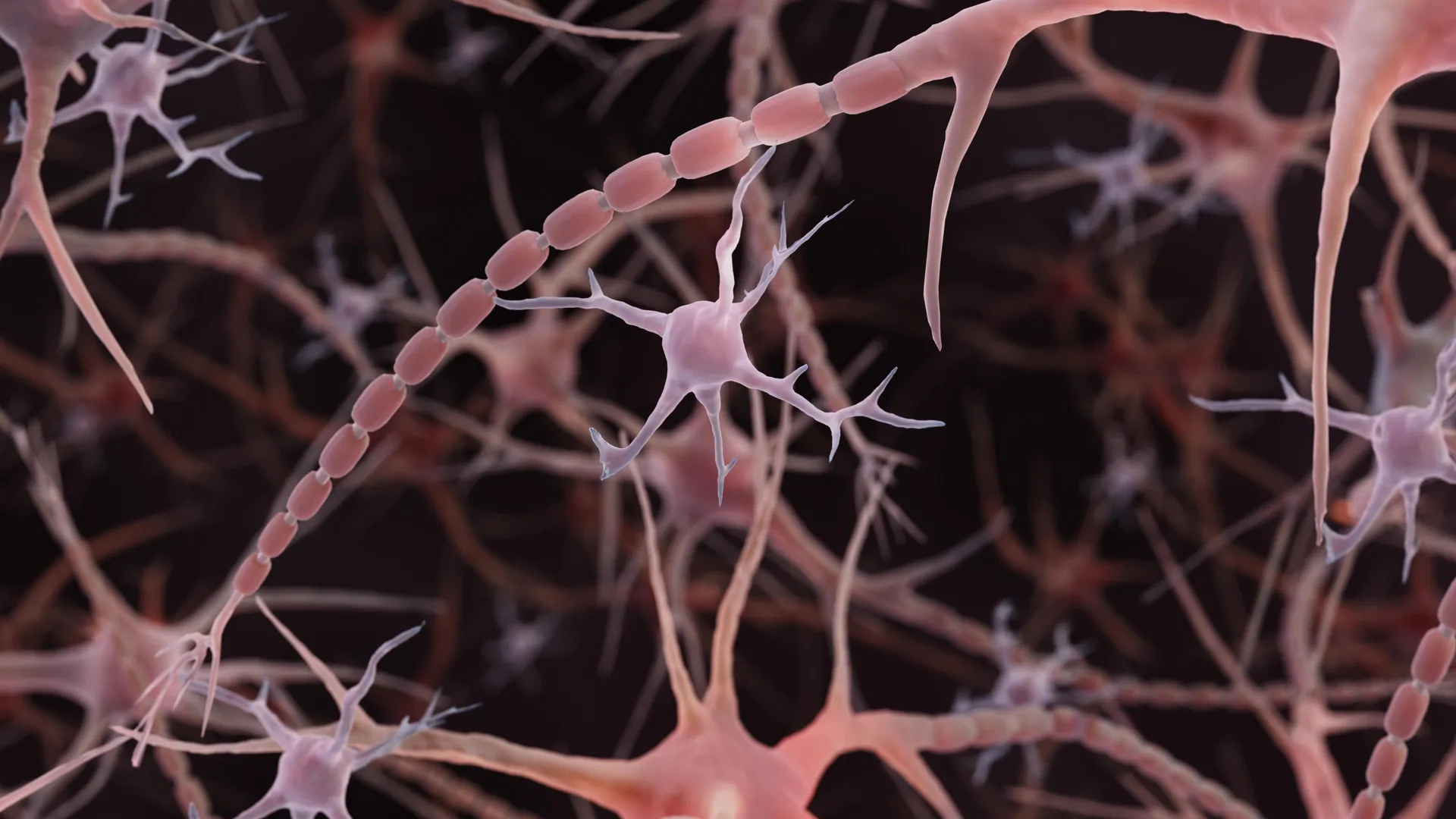
For decades, scientific illustrations and textbooks have favored a simplified model of axons as slender, unblemished conduits. While occasional bulges, known as synaptic varicosities, have been recognized as critical sites for neurotransmitter release and communication with other neurons, the possibility of a more pervasive, nanoscale beading within the axon itself was largely overlooked. This common illustration, though functional for basic conceptualization, may have masked a subtler, yet significant, aspect of neural infrastructure.
The research, published online in Nature Neuroscience on December 2, 2024, utilized advanced imaging techniques to scrutinize mouse neurons under conditions designed to preserve their delicate structures. What emerged was an unexpected and consistent observation: many axons displayed a series of small, repeating swellings, interspersed with narrower segments. These formations, distinct from the larger synaptic varicosities, were termed "non-synaptic varicosities" by the research team, emphasizing their presence outside of established communication junctions.
The implications of this discovery are far-reaching. If these beaded structures are not indicative of damage, as is often the case with pronounced beading in neurodegenerative conditions, but rather a feature of normal axon architecture, then our understanding of how electrical signals propagate needs re-evaluation. The shape and arrangement of these pearls, researchers posit, are not merely incidental but actively influence the speed and efficiency of neuronal signaling.
A Chronology of Discovery: From Worms to Human Tissue
The genesis of this research can be traced back to earlier observations made by Shigeki Watanabe, Ph.D., an associate professor of cell biology and neuroscience at Johns Hopkins University School of Medicine. His initial fascination with repeating pearled structures along axons in simple organisms like worms, sparked by discussions with Swiss scientist Graham Knott, Ph.D., propelled him to investigate this phenomenon further.
A key turning point came with a 2012 study from Harvard University that identified repeating "skeletal" components within axons. Watanabe and Knott hypothesized that if the axon’s internal scaffolding was responsible for the pearling, disrupting this framework might cause the structures to disappear. However, experiments led by Jacqueline Griswold, a Johns Hopkins graduate student and the study’s first author, demonstrated that the pearling persisted even when the internal skeleton was perturbed, redirecting the focus towards the physical properties of the axon’s outer membrane.
This pivot led to a collaboration with Padmini Rangamani, Ph.D., a theoretical biophysicist and professor of pharmacology at the University of California San Diego School of Medicine. Together, they sought to understand how the surrounding membrane could actively shape the axon into this beaded configuration.
Freezing the Moment: Advanced Imaging Techniques Reveal Hidden Details
To capture these exceptionally fine structures, which are approximately 100 times narrower than a human hair, the researchers employed high-pressure freezing electron microscopy. This technique is crucial for preserving the delicate morphology of biological specimens. Unlike conventional electron microscopy, which often involves chemical fixation and dehydration that can distort cellular structures, rapid freezing locks the cell in its near-native state, much like preserving a grape rather than allowing it to desiccate into a raisin.
The team meticulously examined mouse neurons cultured in the laboratory, as well as neurons from adult and embryonic mice. Across tens of thousands of images, the characteristic pearled pattern was consistently observed in nonmyelinated axons – those lacking the insulating myelin sheath. Crucially, further observations using high-resolution imaging of living neurons confirmed that the pearling was not an artifact of the freezing process itself, but a genuine structural feature.
"To see nanoscale structures with standard electron microscopy, we fix and dehydrate the tissues, but freezing them retains their shape — similar to freezing a grape rather than dehydrating it into a raisin," explained Dr. Watanabe in a university announcement. "These findings challenge a century of understanding about axon structure."
The Physics of Form: Membrane Mechanics and Signal Transmission
The researchers then developed mathematical models to elucidate the physical forces at play in creating the beaded axon structure. Their simulations indicated that relatively simple mechanical principles governing the axon’s surrounding membrane could account for the observed pearling, suggesting that a rigid internal mold was not a prerequisite.
Experimental validation followed, reinforcing the role of membrane physics. Manipulating the sugar concentration in the surrounding solution, which affects osmotic pressure and membrane tension, led to observable changes in the size of the swellings. Specifically, increasing sugar concentration, which tends to decrease membrane tension, resulted in smaller bulges.
Further experiments explored the impact of cholesterol, a key component of cell membranes, on axon structure and signaling. When cholesterol was removed, the membrane became less stiff and more fluid. This alteration not only changed the pearled structure in both the models and the mouse neurons but also significantly reduced the speed of electrical signal transmission.
"A wider space in the axons allows ions [chemical particles] to pass through more quickly and avoid traffic jams," Dr. Watanabe elaborated, highlighting the functional consequence of axon morphology. However, the relationship between bead size and signal speed proved complex; the dimensions of both the swellings and the narrow connecting segments were critical determinants of signal velocity.
Activity-Dependent Plasticity: Axons Respond to Electrical Stimulation
Beyond static structure, the research delved into the dynamic nature of axons, investigating whether neural activity could influence their physical form. High-frequency electrical stimulation of the axons resulted in measurable changes: the pearled regions increased in size, becoming, on average, 8% longer and 17% wider. This structural alteration persisted for at least 30 minutes post-stimulation.
Concurrently, electrical signaling within these stimulated axons slowed down, an effect that lasted for at least an hour. Interestingly, when cholesterol had been depleted prior to stimulation, while some structural changes still occurred, the pearling was less pronounced, and the significant slowing of electrical signals was not observed. This suggests that the membrane’s composition and fluidity are integral to the axon’s ability to adapt its structure in response to activity and, consequently, modulate signal transmission. These findings underscore that axons are not simply passive conduits but exhibit a degree of activity-dependent plasticity.
Extending the Vision: Pearled Axons Identified in Human Brain Tissue
The implications of the Johns Hopkins findings were significantly amplified by subsequent research that identified similar pearled axon structures in human brain tissue. This crucial step extended the observations beyond rodent models, providing compelling evidence for the broader relevance of this phenomenon in human neurobiology.
A related study, co-authored by Dr. Watanabe and published online in Neuron on November 24, 2025, and in its February 4, 2026, issue, examined human cortical tissue obtained during epilepsy surgery. Led by Chelsy R. Eddings and her colleagues, this research employed a combination of electrical stimulation and rapid freezing techniques, mirroring the methods used in the earlier mouse studies.
This work not only confirmed the presence of pearled axons in human brain slices but also provided evidence of ultrafast endocytosis, a rapid membrane retrieval process at nerve endings following the release of chemical messengers. While these findings extend the observed phenomenon to human tissue, the researchers caution that not all axons may exhibit this structure uniformly, nor are the signaling effects necessarily identical across the entire human brain. The primary focus of the human study was on membrane recycling at synapses, but the co-occurrence of pearled axons offers a compelling avenue for future investigation.
Broader Implications and Future Directions
The significance of these discoveries has not gone unnoticed by funding agencies and the broader scientific community. The intricate relationship between axon structure and function is now a central focus of a National Institute of Mental Health (NIMH) Multiple Principal Investigator grant awarded to Drs. Watanabe and Rangamani, highlighted in 2024. This initiative supports the development of computational models predicting how a neuron’s physical properties and incoming signals influence its axon, followed by experimental validation in mouse tissues and cell cultures.
Furthermore, independent research is contributing to a growing toolkit for dissecting the complexities of neural wiring. A study published in PLOS Biology on July 17, 2026, by researchers at the University of Edinburgh, utilized automated imaging of living zebrafish to screen 880 compounds for their impact on axon diameter. This screening identified 33 promising compounds, with further confirmation that substances affecting specific chemical signaling pathways could indeed increase axon width. While not a direct replication of the pearling study, this work offers valuable new methods for understanding the regulation of axon size.
For the Johns Hopkins team and the wider neuroscience field, a critical unresolved question remains: how does the pearled structure observed in functioning axons relate to the pronounced beading seen in neurodegenerative diseases like Parkinson’s? Distinguishing between these structurally similar but functionally distinct phenomena is paramount. Understanding this critical difference could enable researchers to better differentiate between normal, adaptive changes in neural wiring and the pathological signs of a system breaking down, paving the way for more targeted diagnostic tools and therapeutic interventions. The implications for treating neurological disorders, from developmental conditions to age-related cognitive decline, are profound, suggesting that subtle architectural features of the brain’s wiring may hold keys to its resilience and vulnerability.