


Researchers at the Kennedy Institute have unearthed compelling evidence suggesting that certain joints may possess an inherent vulnerability to inflammatory arthritis even before birth. This groundbreaking discovery has the potential to illuminate a persistent enigma within the study of rheumatoid arthritis (RA): the disease’s uncanny predilection for specific joints while leaving others remarkably unscathed. The findings, published in the prestigious journal Nature Immunology, propose that this site-specific attack is not solely dictated by the immune system, but also by fundamental biological distinctions established within individual joints during their developmental stages.
Unraveling the Mystery of Joint Selectivity in Rheumatoid Arthritis
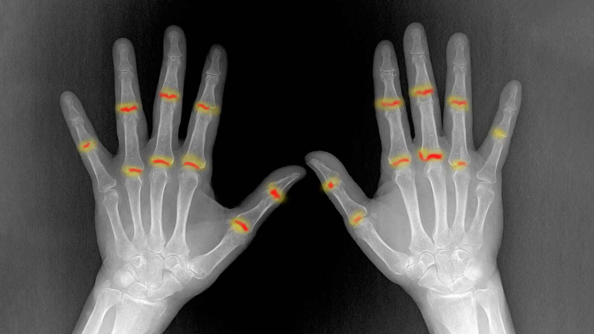
Rheumatoid arthritis, a chronic autoimmune condition, is characterized by the immune system mistakenly targeting the synovium – the delicate tissue lining the joints. This aberrant immune response triggers a cascade of debilitating symptoms, including pain, swelling, and stiffness. Over time, this inflammation can inflict significant damage on the cartilage, bone, and surrounding connective tissues, leading to progressive joint deterioration and loss of function. For decades, clinicians and scientists have observed that RA disproportionately affects certain joints, most commonly the proximal interphalangeal (PIP) joints of the fingers, while distal interphalangeal (DIP) joints, located closer to the fingertips, are often spared. This observation has fueled a long-standing quest to understand the underlying mechanisms driving this selective vulnerability.
The study, titled "The embryonic origins of site-specific arthritis," meticulously investigated the differences between PIP and DIP joints, which exhibit starkly contrasting patterns of disease susceptibility in RA. By employing advanced techniques such as single-cell sequencing, sophisticated image analysis, and high-resolution 3D X-ray scanning, the research team constructed a detailed developmental map of human finger joints. This allowed them to scrutinize the nascent stages of joint formation, a period when the fundamental architectural and cellular blueprints are laid down.
Pre-Natal Blueprint: Identifying Early Joint Distinctions
A pivotal finding of the research is the identification of significant biological differences between PIP and DIP joints that are already present before birth. The researchers observed that PIP joints, the ones frequently targeted by RA, possess a larger volume of synovial tissue compared to DIP joints. Furthermore, these vulnerable joints were found to contain a greater abundance of PI16-positive (PI16+) fibroblasts. Fibroblasts are specialized cells within connective tissue that play a crucial role in tissue repair and maintenance. The presence of these PI16+ fibroblasts in higher numbers within PIP joints during embryonic development suggests that the local tissue environment of a joint may be a critical determinant of its susceptibility to inflammation later in life.
Professor Christopher Buckley, Kennedy Professor of Translational Rheumatology at the University of Oxford and a senior author on the study, commented on the significance of these findings. "For decades, we have known that rheumatoid arthritis selectively targets particular joints, but one of the great unanswered questions is why?" he stated. "Our findings suggest that the answer lies not only in the immune system but also in the tissues themselves. The cellular and structural characteristics established during development may help determine where inflammation takes hold later in life." This statement underscores a paradigm shift in understanding RA, moving beyond a purely immune-centric view to incorporate developmental biology.
Mapping the Architecture of Developing Joints
The research team’s meticulous mapping of developing human joints revealed that in their early stages, these structures are predominantly composed of structural cells, such as fibroblasts and cartilage-forming cells, rather than immune cells. The study delved into the signaling pathways that guide the differentiation of these progenitor cells into specialized types. A particular focus was placed on the fibroblasts that ultimately form the synovial lining. These cells are vital for joint health, producing lubricating substances that facilitate smooth movement and protect the joint. However, in the context of arthritis, these same cells can exhibit aberrant behavior.
Further analysis indicated that the synovial lining can originate from multiple sources, including cartilage and surrounding joint fibroblasts. The local environmental conditions, such as areas of low oxygen, were also found to influence this developmental process. Understanding these intricate developmental signals could offer invaluable insights into how synovial fibroblasts acquire their specific functions and, crucially, might reveal potential avenues for restoring their normal protective roles in the context of arthritis.
Specialized Cellular Signatures in Arthritis-Prone Joints
The study identified several key distinctions between PIP and DIP joints at the cellular level. Employing a specially developed image analysis tool, the researchers demonstrated that PI16+ fibroblasts were significantly more prevalent in PIP joints. These cells were often found concentrated in areas surrounding blood vessels and at the critical junctures where tendons and ligaments connect to adjacent tissues.
Crucially, PI16+ fibroblasts exhibited differential responses to inflammatory signals compared to other fibroblast populations. While both PI16+ and PI16- fibroblasts generally displayed a pro-inflammatory reaction, the PI16+ cells showed distinct alterations in biological pathways involved in immune regulation and tissue organization. These differences were not confined to individual cell types; high-resolution 3D imaging conducted at the Diamond Light Source revealed that PIP joints possessed a greater volume of synovial tissue, and this tissue was structured differently compared to the synovium in joints typically spared by RA. These combined cellular and structural variations provide a compelling explanation for the increased propensity of certain joints to develop inflammation.
Dr. Sarah Davidson, a Postdoctoral Researcher at the Kennedy Institute and one of the study’s first authors, highlighted the implications of these cellular differences. "We found that joints commonly affected by rheumatoid arthritis already contain distinct cellular populations before birth," she explained. "PI16+ fibroblasts were enriched in vulnerable joints and responded differently to inflammatory signals. Their location and behavior suggest they could help shape where disease develops."
A Developmental Perspective on Rheumatoid Arthritis Susceptibility
The research’s findings collectively propose a more comprehensive explanation for the site-specific nature of rheumatoid arthritis. Instead of joint vulnerability being solely determined by immune system activity later in life, the propensity for inflammation to take hold appears to be influenced by cellular and structural characteristics established during the very formation of the joints. This implies that the inherent biological environment of each joint, shaped during its embryonic development, plays a significant role in predisposing it to rheumatoid arthritis years, or even decades, later.
Broader Implications and Future Directions
This discovery opens up exciting new avenues for research and potential therapeutic strategies for rheumatoid arthritis. If specific developmental pathways and cellular characteristics confer vulnerability, then interventions targeting these early processes, or even modifying the environment of developing joints, could theoretically be explored. While such interventions are likely many years away, this research provides a crucial foundation for future studies aimed at understanding and preventing this debilitating disease.
The implications extend beyond RA, potentially offering insights into other inflammatory conditions that exhibit site-specific patterns. Understanding the interplay between developmental biology and immune responses in joint health could lead to novel diagnostic tools that identify individuals at higher risk of developing RA, allowing for earlier intervention and management. Furthermore, pinpointing the precise molecular mechanisms by which PI16+ fibroblasts contribute to inflammation could pave the way for the development of targeted therapies that specifically modulate the behavior of these cells, thereby preventing or mitigating joint damage.
The collaborative nature of this research, involving scientists from the Kennedy Institute of Rheumatology at the University of Oxford, the University of Birmingham, University College London, and Diamond Light Source, underscores the multidisciplinary approach required to tackle complex biological questions. Supported by the Medical Research Council (MRC), this study represents a significant leap forward in our understanding of rheumatoid arthritis, moving us closer to unraveling its long-standing mysteries and, ultimately, to developing more effective strategies for its prevention and treatment. The chronological progression of this research, from initial observation of RA’s predilection for certain joints to the detailed mapping of embryonic development, highlights the incremental yet powerful advances in scientific inquiry. The data gathered, particularly the detailed cellular and structural comparisons between PIP and DIP joints, provides robust evidence for the developmental origins of disease susceptibility, offering a beacon of hope for future breakthroughs in rheumatology.