


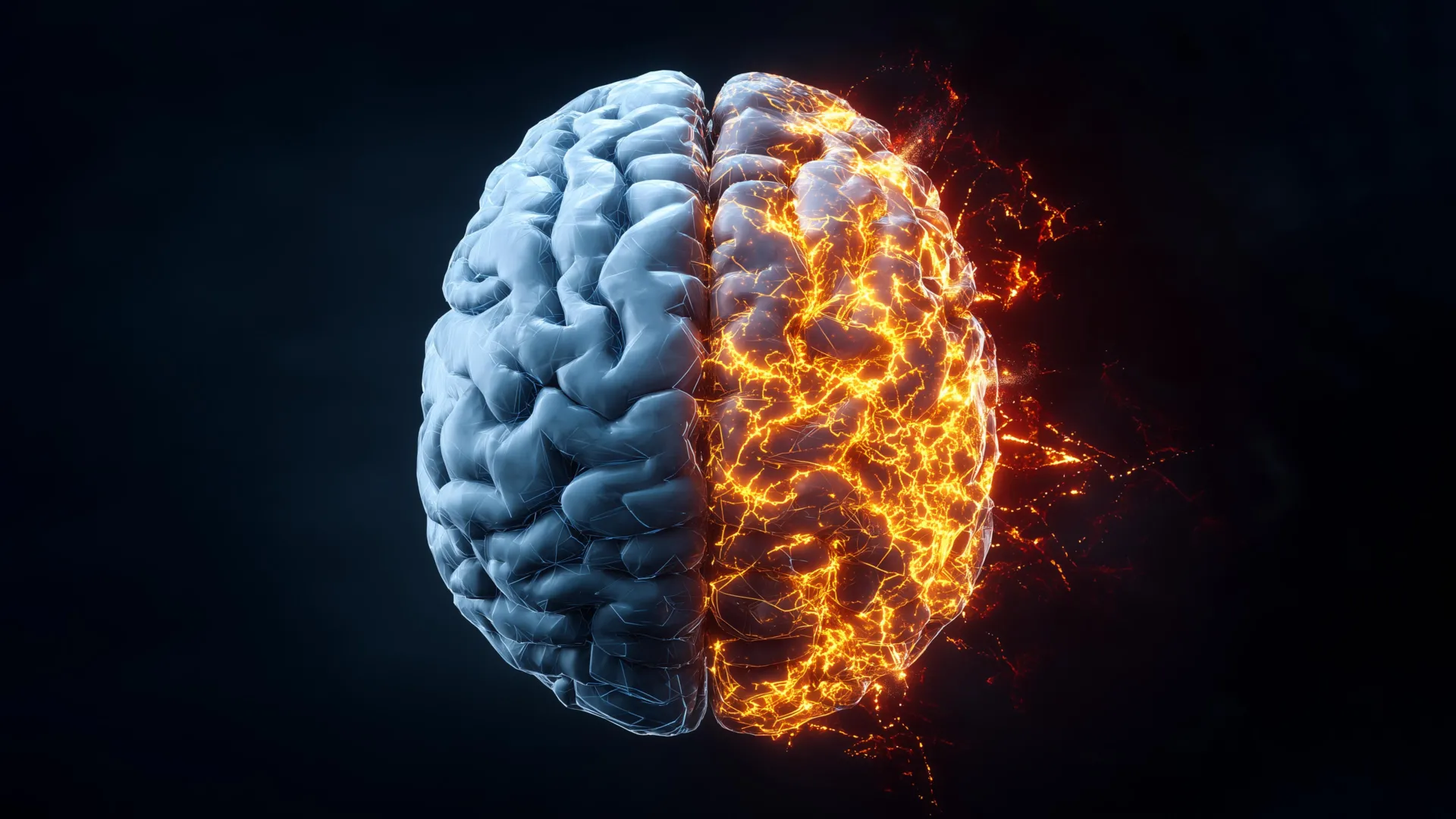
Researchers at Rice University have achieved a groundbreaking milestone in Alzheimer’s research, creating the first comprehensive, label-free molecular atlas of the disease in an animal model. This pioneering work provides an unprecedentedly detailed view of how Alzheimer’s disease originates and progresses, offering critical insights into the complex biochemical landscape of the affected brain. Alzheimer’s disease, a devastating neurodegenerative condition that claims more lives annually than breast and prostate cancers combined, presents a profound public health challenge, making understanding its underlying mechanisms an urgent priority for scientists worldwide.
The innovative approach, detailed in the latest issue of ACS Applied Materials and Interfaces, merges an advanced light-based imaging technique with sophisticated machine learning algorithms. By examining brain tissue from both healthy and Alzheimer’s-affected animals, the Rice team has revealed that the chemical alterations characteristic of Alzheimer’s extend far beyond the well-known amyloid plaques. Instead, these changes are distributed unevenly and intricately throughout the brain, painting a more complex picture of the disease’s pathology than previously understood.
Advanced Laser Imaging Illuminates Brain Chemistry with Unprecedented Detail
To capture these subtle yet significant chemical shifts, the scientists employed hyperspectral Raman imaging, a highly advanced form of Raman spectroscopy. This technique utilizes a precisely tuned laser to excite molecules within the tissue, causing them to emit light at specific wavelengths. These emissions act as unique "chemical fingerprints," allowing researchers to identify and quantify the presence and distribution of various chemical compounds.
"Traditional Raman spectroscopy provides a single data point of chemical information at a specific molecular site," explained Ziyang Wang, an electrical and computer engineering doctoral student at Rice and a first author on the study. "Hyperspectral Raman imaging, however, is akin to taking thousands of these measurements across an entire tissue slice. This allows us to construct a comprehensive molecular map, revealing the intricate variations in chemical composition across different regions of the brain."
The research team meticulously scanned entire brain slices, accumulating vast numbers of overlapping measurements. This extensive data collection enabled the construction of high-resolution molecular maps, depicting the chemical makeup of both healthy and diseased brain tissue with remarkable clarity. A key advantage of this methodology is its "label-free" nature. Unlike conventional techniques that require samples to be treated with dyes, fluorescent proteins, or molecular tags, this method observes the tissue in its natural state.
"This means we were able to observe the brain as it is, without any artificial modifications," Wang emphasized. "This approach provides a complete, unaltered portrait of its chemical composition. We believe this unbiased perspective is crucial for discovering novel disease-related changes that might otherwise be overlooked by methods requiring sample labeling." The absence of labels also eliminates potential artifacts or interactions that could skew experimental results, ensuring a more accurate representation of the biological reality.
Machine Learning Deciphers the Complex and Uneven Spread of Alzheimer’s Damage
The sheer volume of data generated by the hyperspectral Raman imaging process necessitated the use of powerful analytical tools. The researchers turned to machine learning (ML) to process and interpret these extensive datasets. Initially, they applied unsupervised ML algorithms. These algorithms were designed to identify inherent patterns within the chemical signals without any prior assumptions about what those patterns might represent. This initial analysis effectively sorted tissue samples based solely on their unique molecular characteristics, allowing the data itself to guide the discovery process.
Following this exploratory phase, the team employed supervised ML. In this stage, the algorithms were trained to differentiate between samples from healthy brains and those affected by Alzheimer’s disease. This training allowed the researchers to quantify how strongly different brain regions exhibited Alzheimer’s-related chemistry, providing a detailed understanding of the disease’s spatial impact.
"Our analysis revealed a critical finding: the chemical changes associated with Alzheimer’s disease are not uniformly distributed throughout the brain," stated Wang. "Certain brain regions display significant chemical alterations, while others remain comparatively less affected. This uneven distribution pattern offers valuable insights into why Alzheimer’s symptoms often emerge gradually and why therapeutic strategies that target a single pathological aspect have historically encountered limited success." This nuanced understanding of disease progression could revolutionize the development of more targeted and effective treatments.
Uncovering Metabolic Disruptions in Crucial Memory Regions
Beyond the accumulation and misfolding of proteins, the study unveiled broader metabolic discrepancies between healthy and Alzheimer’s-affected brains. The levels of key biomolecules, including cholesterol and glycogen, were found to vary significantly across different brain regions. The most pronounced differences were observed in areas critically involved in memory formation and retrieval, specifically the hippocampus and the cortex.
Cholesterol plays a vital role in maintaining the structural integrity of brain cells and is essential for proper neuronal function. Glycogen, on the other hand, serves as a readily available source of energy for brain cells, particularly during periods of high metabolic demand.
"Cholesterol is fundamental for maintaining brain cell structure, and glycogen acts as a localized energy reserve," explained Shengxi Huang, an associate professor of electrical and computer engineering and materials science and nanoengineering at Rice, and the corresponding author of the study. "Taken together, these findings strongly support the growing understanding that Alzheimer’s disease involves widespread disruptions in brain structure and energy balance, rather than being solely attributable to protein aggregation and misfolding." Huang’s affiliations also include the Ken Kennedy Institute, the Rice Advanced Materials Institute, and the Smalley-Curl Institute, highlighting the interdisciplinary nature of this research.
This discovery of metabolic dysregulation in critical memory centers suggests that Alzheimer’s may impair the brain’s ability to maintain its structural integrity and efficiently manage its energy resources, contributing to the cognitive decline characteristic of the disease. The implications for understanding disease mechanisms and developing interventions are substantial, pointing towards the need for approaches that address these broader metabolic vulnerabilities.
A Broader Perspective on Alzheimer’s Progression: From Small Areas to Whole-Brain Mapping
The genesis of this extensive research project can be traced back to ongoing discussions among scientists at Rice about novel methodologies for studying the complexities of the Alzheimer’s brain. Initially, the team’s investigations were confined to analyzing relatively small areas of brain tissue. However, a pivotal shift in perspective emerged.
"At first, our focus was on measuring only limited sections of brain tissue," Wang recalled. "Then, the question arose: what if we could map the entire brain and gain a much more comprehensive overview of the disease’s molecular landscape? It required numerous iterations of testing and experimentation before we could effectively integrate the measurement and analysis components." This iterative process, characterized by trial and error, was essential for refining the hyperspectral Raman imaging technique and the subsequent machine learning protocols to a point where they could reliably generate and interpret whole-brain molecular data.
The moment when the complete chemical map of the Alzheimer’s brain finally coalesced was met with significant impact. New patterns, previously invisible to conventional imaging techniques, began to emerge.
"Patterns appeared that had not been visible under regular imaging," Wang stated, reflecting on the profound discovery. "Witnessing those results was deeply satisfying. It felt akin to uncovering a hidden layer of information that had been present all along, awaiting the appropriate analytical approach to reveal its secrets." This metaphor underscores the transformative power of the new methodology, which has effectively unveiled previously inaccessible aspects of the disease’s molecular underpinnings.
Implications for Diagnosis and Therapeutic Development
By providing the first detailed, label-free chemical maps of the Alzheimer’s brain, this groundbreaking research offers a more holistic and accurate understanding of the disease’s progression. The ability to visualize the intricate and uneven distribution of chemical changes throughout the brain, independent of artificial labels, provides a powerful new tool for researchers.
The team expresses optimism that these findings will pave the way for earlier and more accurate diagnosis of Alzheimer’s disease. By identifying specific molecular signatures and their spatial distribution, it may become possible to detect the disease at its nascent stages, even before the onset of overt clinical symptoms. Furthermore, a deeper comprehension of the diverse and uneven molecular alterations could lead to the development of more sophisticated and effective therapeutic strategies. Instead of a one-size-fits-all approach, future treatments might be designed to address specific molecular pathways or target particular brain regions that are disproportionately affected by the disease.
The research was generously supported by grants from the National Science Foundation (awards 2246564 and 1934977), the National Institutes of Health (grant 1R01AG077016), and the Welch Foundation (award C2144), underscoring the national and institutional commitment to advancing Alzheimer’s research. The collaborative efforts and substantial funding have been instrumental in enabling this significant leap forward in understanding one of the most pressing health challenges of our time. The scientific community will undoubtedly be watching closely as this research continues to unfold, with the hope that it will ultimately contribute to alleviating the burden of Alzheimer’s disease for millions worldwide.