


The human body’s intricate choreography of life is orchestrated by a meticulously organized set of genetic instructions, dictating the growth and function of every cell. Cancer, a devastating disease, often begins when these fundamental instructions become corrupted. Over time, cells can accumulate a cascade of genetic errors, liberating them from the body’s natural growth restraints and paving the way for uncontrolled proliferation. Among the earliest telltale signs of this insidious process are chromosomal abnormalities – deviations in the number or structure of chromosomes. These defects act as critical triggers, nudging otherwise healthy cells down the path toward malignancy.
Groundbreaking AI Technology Illuminates Early Cancer Mechanisms
In a significant leap forward for cancer research, scientists at the European Molecular Biology Laboratory (EMBL) in Heidelberg have unveiled a powerful artificial intelligence (AI)-driven tool. This innovative technology is poised to revolutionize how researchers investigate the genesis of chromosomal abnormalities, offering unprecedented insights into the conditions that foster these errors. By shedding light on the very origins of these cellular missteps, the EMBL team’s development holds immense promise for a deeper understanding of how cancer initiates and progresses.
Jan Korbel, a senior scientist at EMBL and senior author of the groundbreaking study published in the prestigious journal Nature, emphasized the critical role of chromosomal aberrations in aggressive cancers. "Chromosomal abnormalities are a main driver for particularly aggressive cancers, and they’re highly linked to patient death, metastasis, recurrence, chemotherapy resistance, and fast tumor onset," Korbel stated. "We wanted to understand what determines the likelihood that cells undergo such chromosomal alterations, and what’s the rate at which such abnormalities arise when a still normal cell divides."
A Century-Old Theory Validated by Modern Technology
The suspicion that faulty chromosomes might be linked to cancer is not a new one; it dates back over a century. In the early 20th century, German scientist Theodor Boveri, a pioneer in cell biology, first proposed this revolutionary idea. His meticulous observations under the microscope led him to theorize that abnormal chromosomal content within cells could be a key factor in cancer development. Boveri’s hypothesis, though remarkably prescient, remained largely theoretical for decades due to the immense technical challenges involved in its empirical validation.
Historically, studying these chromosomal anomalies has been a formidable undertaking. At any given moment, only a small fraction of cells within a sample exhibit visible chromosomal defects. Furthermore, many of these compromised cells are naturally eliminated through cellular selection processes or undergo programmed cell death. This scarcity made traditional research methods incredibly labor-intensive. Scientists would painstakingly search for these rare aberrant cells manually under a microscope, a process that allowed for the isolation of only a handful of cells for subsequent, in-depth analysis. This bottleneck severely limited the scope and scale of investigations into the fundamental mechanisms of cancer initiation.
The Genesis of MAGIC: A Collaborative Effort
The impetus for developing a more efficient solution came from Marco Cosenza, a Research Scientist in the Korbel Group. Cosenza recognized the shared technical hurdles faced by various EMBL teams investigating cellular processes. Through fruitful collaborations with colleagues across EMBL, he played a pivotal role in designing an automated platform that ingeniously integrates advanced microscopy, single-cell sequencing, and cutting-edge artificial intelligence. This powerful system, christened machine learning-assisted genomics and imaging convergence (MAGIC), represents a paradigm shift in biological research.
The development of MAGIC can be traced back to the initial research phase where scientists manually identified cells with specific visual markers indicative of chromosomal instability. This laborious process involved hours spent examining microscope slides. The limitations of this manual approach became increasingly apparent as the volume of data required for robust statistical analysis grew. The desire to accelerate discovery and expand the investigable cell population fueled the drive to automate this critical identification step. The COVID-19 pandemic, while presenting significant challenges, also provided an unexpected opportunity for Cosenza to dedicate focused time to exploring AI-driven solutions. "This project combined a lot of my interests in one," Cosenza reflected. "It involves genomics, microscopic imaging, and robotic automation. During the COVID-19-related lockdown in 2020, I could really spend some time on learning and applying AI computer vision technologies to the biological image data we had collected before. Afterwards, we designed experiments to validate it and take it further."
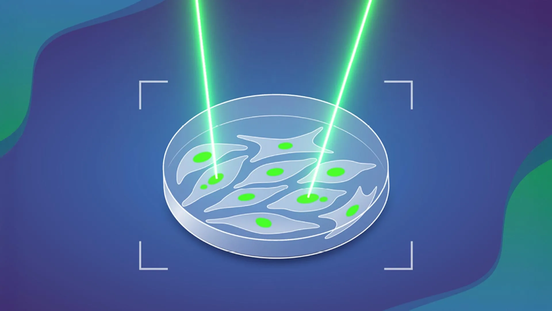
MAGIC: An AI-Powered Cellular "Laser Tag" System
The MAGIC system operates with a remarkable degree of automation, akin to a sophisticated, high-throughput cellular identification system. It efficiently scans through vast populations of cells, pinpointing those that exhibit a specific, visually discernible characteristic. For this particular study, the researchers focused their attention on a structure known as a ‘micronucleus’.
Micronuclei are small, membrane-bound vesicles that form within the cytoplasm of a cell. They contain fragments of DNA that have become separated from the main genome during cell division. The presence of micronuclei is a strong indicator of underlying chromosomal instability and is strongly associated with an increased likelihood of developing further chromosomal abnormalities, thereby elevating the risk of a cell becoming cancerous.
Once MAGIC detects a cell containing a micronucleus, it precisely marks that cell using a laser. This sophisticated tagging mechanism relies on a specialized photoconvertible dye. This fluorescent molecule possesses the unique property of altering the color of light it emits upon exposure to specific wavelengths of light, effectively creating a permanent, identifiable tag on the targeted cell. This targeted labeling ensures that researchers can reliably isolate the cells of interest from a complex cellular mixture for subsequent, more detailed investigations.
The Automated Workflow of the MAGIC System
The operational sequence of the MAGIC system is elegantly designed for efficiency and precision. The process commences with an automated microscope systematically capturing a comprehensive array of images from a given cell sample. Following image acquisition, a sophisticated machine learning algorithm, meticulously trained on a large dataset of manually labeled images of micronucleus-containing cells, undertakes the critical task of image analysis. This algorithm is capable of discerning subtle visual cues that differentiate micronucleus-bearing cells from their normal counterparts with remarkable accuracy.
Upon identifying a cell that contains a micronucleus, the algorithm instantly relays the precise coordinates of that cell to the microscope. The microscope then precisely directs a focused beam of light onto the designated cell, activating the photoconvertible dye and permanently tagging the cell. These meticulously tagged cells can subsequently be isolated from the living cell population using established techniques such as flow cytometry. Once isolated, these highly enriched populations of potentially aberrant cells are available for in-depth molecular analysis, including comprehensive genomic sequencing.
By effectively automating the laborious and time-consuming manual search for micronuclei, MAGIC empowers scientists to examine a vastly larger number of cells than was previously feasible. In a remarkably short timeframe, often less than a single day, the system can meticulously analyze close to 100,000 individual cells. This dramatic increase in throughput significantly accelerates the pace of discovery and allows for the identification of rare cellular events that might otherwise be missed.
Quantifying the Spontaneous Occurrence of Chromosomal Errors
Leveraging the power of MAGIC, the EMBL researchers embarked on a comprehensive study of chromosomal abnormalities within cultured human cells. These cells were meticulously derived from healthy, normal human cell lines, providing a pristine baseline for their investigations. Their analysis yielded a striking quantitative insight: a substantial proportion, slightly more than 10%, of cell divisions in these normal cells spontaneously give rise to chromosomal abnormalities. This finding underscores the inherent fragility of the genome and the constant cellular challenges that must be overcome to maintain genetic integrity.
The study further revealed a significant amplification of this rate when a critical tumor suppressor gene, p53, is mutated. The p53 gene is a well-known guardian of the genome, playing a crucial role in preventing the proliferation of damaged cells. When p53 is dysfunctional, the rate at which spontaneous chromosomal abnormalities occur nearly doubles, highlighting the pivotal role of this gene in maintaining genomic stability and preventing cancer development. This nearly 100% increase in the rate of abnormalities in p53-mutated cells provides crucial quantitative data for understanding the accelerated progression towards cancer in such scenarios.
Beyond the influence of p53, the research team also delved into other factors that can modulate the formation of chromosomal abnormalities. Their investigations examined the impact of double-stranded DNA breaks within chromosomes, including their presence and precise location. Such breaks are known to be highly mutagenic events that can lead to chromosomal rearrangements and numerical aberrations, further contributing to genomic instability.
Broad Implications for Biological Discovery
The success of this pioneering research was significantly bolstered by extensive collaborations, both within EMBL and with external institutions. Key contributions came from the Advanced Light Microscopy Facility (ALMF) and the Pepperkok Team at EMBL Heidelberg, who provided essential expertise in advanced imaging techniques. Isidro Cortes-Ciriano’s group at EMBL-European Bioinformatics Institute (EMBL-EBI) offered crucial bioinformatics support, while Andreas Kulozik’s team at the German Cancer Research Centre (DKFZ), part of the Molecular Medicine Partnership Unit (MMPU) between EMBL and Heidelberg University, provided valuable clinical and biological context. This interdisciplinary approach highlights the collaborative spirit driving modern scientific advancement.
The inherent flexibility and adaptability of the MAGIC system are key to its broad potential. While the current study focused on training the AI to identify micronuclei, the underlying machine learning architecture is capable of being retrained to recognize a diverse array of other cellular features. "As long as you have a feature that can be discriminated visually from a ‘regular’ cell, you can — thanks to AI — train the system to detect it," Korbel explained. This modularity means that MAGIC can be readily deployed to address a multitude of research questions across various biological disciplines.
The implications of this AI-powered platform extend far beyond cancer research. It holds the potential to accelerate discoveries in developmental biology, immunology, infectious diseases, and fundamental cell biology. By providing a robust and efficient method for identifying and isolating rare cellular events, MAGIC promises to unlock new avenues of investigation and deepen our understanding of the complex processes that govern life at the cellular level. The ability to rapidly screen large cell populations for specific morphological or structural markers could revolutionize fields such as drug screening, where identifying rare responder cells is critical, or in the study of stem cell differentiation, where subtle cellular changes are paramount. The quantitative data generated by MAGIC regarding the rate of spontaneous errors, particularly in the context of gene mutations, also provides a crucial baseline for understanding the long-term effects of environmental exposures and genetic predispositions on cellular health. The development represents a significant stride in making complex biological research more accessible and accelerating the translation of fundamental discoveries into tangible benefits for human health.